Issue 59
Aug 2026
AFFAIRS OF THE HEART
By Dr Sia Ching Hui, Consultant, National University Heart Centre Singapore, Adjunct Assistant Professor, Department of Medicine, NUS Yong Loo Lin School of Medicine
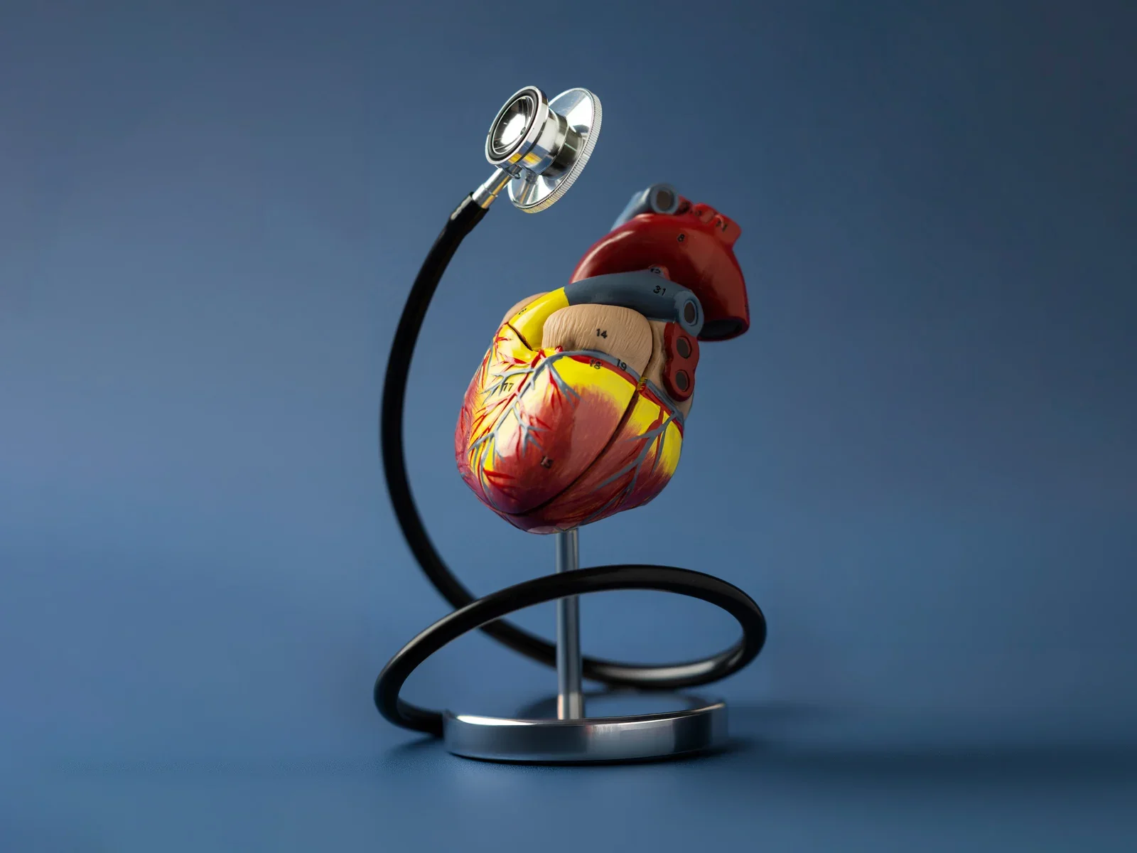
How multimodality cardiac imaging is transforming cardiovascular care and research.
When a patient presents with chest pain, breathlessness or the sequelae of a stroke, the immediate clinical question is: what is the heart doing, and why? For much of the twentieth century, answering that question required catheters threaded into the heart, needles into the myocardium, and patients exposed to the risks of invasive procedures. Today, on the National University Health System (NUHS) campus, we are leading a transformation—the full depth of cardiac biology can be interrogated non-invasively, with cameras and magnets and intelligent algorithms, without a single incision.
Four modalities, one heart
Modern multimodality cardiac imaging deploys four complementary technologies. Echocardiography (cardiac ultrasound) is our most accessible workhorse: affordable, radiation-free, and available at the bedside via portable devices. Computed tomography (CT) generates high-resolution three-dimensional images of the coronary arteries and cardiac structures: it yields anatomical and functional information once obtainable only in the catheterisation laboratory. Cardiac Magnetic Resonance Imaging (CMR) characterises the myocardium at the tissue level, quantifying fibrosis, inflammation, oedema and perfusion without ionising radiation. Finally, nuclear imaging like Positron Emission Tomography (PET) deploys radiolabelled tracers to measure molecular-level processes—inflammation, receptor expression and metabolic activity.
We operate all four modalities including cutting-edge hybrid platforms such as simultaneous PET/MRI and PET/CT acquisitions, which co-register molecular and structural cardiac data in a single examination. Each modality answers questions that guide real decisions: whether to operate, which medication to prescribe, whether patient’s risk justifies intervention and, increasingly, whether an invasive test is needed at all.
Non-invasive evaluation: Replacing the catheter and the biopsy
One of the most consequential contributions of modern cardiac imaging is its growing ability to replace invasive testing. Consider coronary physiology: traditionally, determining whether a coronary narrowing is haemodynamically significant required inserting a pressure wire directly into the artery during an invasive angiogram. CT-derived fractional flow reserve (CT-FFR) now achieves this from a standard cardiac CT scan, using computational fluid dynamics to model coronary blood flow and sparing thousands of patients an invasive procedure while delivering equivalent diagnostic accuracy.
The same approach and benefit apply at the molecular level. Understanding processes relating to cardiac hypertrophy and fibrosis has historically required invasive myocardial biopsy. Our clinical group at the National University Heart Centre Singapore (NUHCS) is pioneering non‐invasive fibrosis imaging using cutting-edge PET radiotracers. We are studying ischaemic and hypertrophic cardiomyopathy patients to understand fibrosis patterns across the heart, correlating findings with CMR-derived parameters of ejection fraction, myocardial strain, mass and fibrosis. If validated, this allows clinicians to non‐invasively phenotype patients’ molecular cardiac biology and monitor the effects of novel anti‐fibrotic therapies. This is a window into cardiac disease that biopsy cannot provide at scale.
CT also enables non-invasive interrogation of coronary plaque biology. Following acute myocardial infarction (AMI) in patients, our collaborative work has demonstrated that post-AMI coronary plaques display heightened noncalcified components, particularly fibrofatty tissue and necrotic core, alongside increased pericoronary adipose tissue (PVAT) attenuation—a marker of vascular inflammation, and all detectable on CT without touching the vessel wall. The NUHCS now has access to a next-generation photon counting CT machine, which will enable older patients and patients with coronary stents to be imaged while also allowing our team to characterise the myocardium. This refines risk stratification in ways that conventional angiography cannot.
One of the most consequential contributions of modern cardiac imaging is its growing ability to replace invasive testing. If validated, this allows clinicians to non‐invasively phenotype patients’ molecular cardiac biology and monitor the effects of novel anti‐fibrotic therapies. This is a window into cardiac disease that biopsy cannot provide at scale.”
Advanced acquisition: 4D flow, total-body PET and the mechanistic frontier
Beyond replacing invasive tests, our programme is developing new acquisition paradigms that expand what imaging can reveal. Four-dimensional flow MRI (4D flow CMR) encodes blood velocity in all three spatial dimensions throughout the cardiac cycle, enabling precise quantification of intracardiac blood stasis, turbulent kinetic energy and flow inefficiency. We are applying this to patients with complex heart diseases, seeking to establish whether abnormal flow dynamics predict worse outcomes.
Total-body PET/CT represents another leap in acquisition capability. We can simultaneously image the entire body in a single examination. We can image the entire body’s organs and vascular bed in a single scan and also examine biological processes simultaneously.
Imaging as a lens on treatment: Drugs, surgery and lifestyle
A major strength of cardiac imaging is its power to make therapeutic change visible. We have used echocardiography and CMR to evaluate the cardiac impact of emerging treatments. In bariatric surgery for example, imaging of myocardial strain, a sensitive marker of subtle contractile dysfunction that precedes any fall in ejection fraction, has shown that surgical weight loss significantly improves both left ventricular and left atrial strain, effects invisible to standard functional measures. These findings position imaging as an indispensable endpoint for understanding how treatments work and who benefits most.
Cardiovascular imaging is not, as is sometimes assumed, just about producing pretty pictures. It is the cornerstone of diagnosis, the lens through which we understand disease mechanisms, the surrogate endpoint that makes clinical trials possible, and increasingly, the platform upon which AI will transform the speed, accuracy, and accessibility of cardiac care.”
The heart-brain axis: A new research frontier
The most compelling illustration of multimodality imaging’s power is its application to the heart-brain axis. Cardiovascular and cerebrovascular diseases are profoundly interlinked: poor cardiac function causes chronic brain hypoperfusion, atrial fibrillation raises both stroke and dementia risk, and acute stroke itself triggers neurogenic cardiac injury. Through our research programme, led jointly by cardiologists and stroke neurologists, we are using the full imaging arsenal to interrogate these connections. In cryptogenic stroke, where approximately 20% of ischaemic strokes have no identified cause, we deploy combined PET/MRI with novel fibrosis and inflammation tracers to unmask occult heart disease invisible to standard investigations.
Artificial Intelligence (AI): Democratising the image
Cardiac imaging has a structural vulnerability: it is operator-dependent, training-intensive and prone to human variability. AI addresses this directly. We performed an AI-Echo trial to evaluate whether an AI-powered point-of-care echocardiographic platform can detect cancer therapy-related cardiac dysfunction in HER2-positive breast cancer patients earlier and more accurately than standard echocardiography. Preliminary results show the AI classifies left ventricular ejection fraction with over 90% accuracy against expert readers. The implications extend far beyond oncology: a clinical coordinator, guided by AI, obtaining reliable cardiac measurements at the bedside is an emerging reality that could democratise imaging access across resource-limited settings.
Conclusions
The work described here is built on multidisciplinary collaboration between cardiologists, radiologists, nuclear medicine physicians, biomedical engineers and AI researchers. Together, we are building a richly phenotyped cardiac imaging dataset unique to Asia.
Cardiovascular imaging is not, as is sometimes assumed, just about producing pretty pictures. It is the cornerstone of diagnosis, the lens through which we understand disease mechanisms, the surrogate endpoint that makes clinical trials possible, and increasingly, the platform upon which AI will transform the speed, accuracy, and accessibility of cardiac care.
More from this issue