Equipment
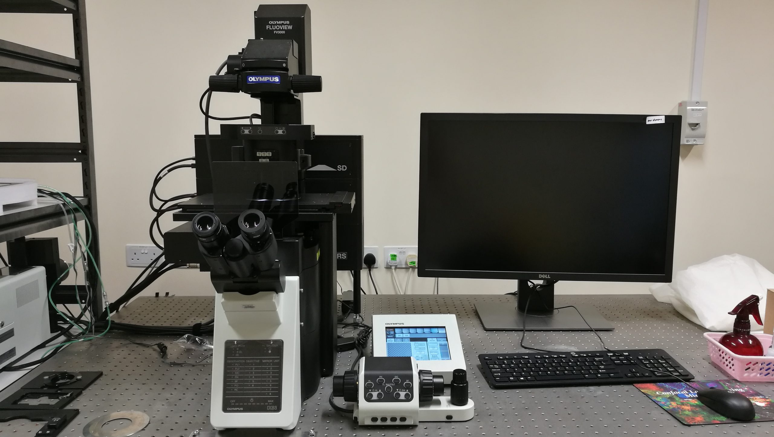
Olympus FV3000 Confocal Microscope
Lasers: 405nm, 488nm, 561nm, 640nm
Applications:
- High resolution and High sensitivity Imaging
- Z-stack
- Time lapse (Z-drift compensation and Multi-position capability)
- Co-localization
- Fluorescent Resonant Energy Transfer (FRET)
- Fluorescent Recovery after Photo-bleaching (FRAP)
- Macro to Micro
- Stitching
- Confocal Reflectance Imaging
- Multichannel Spectral Imaging
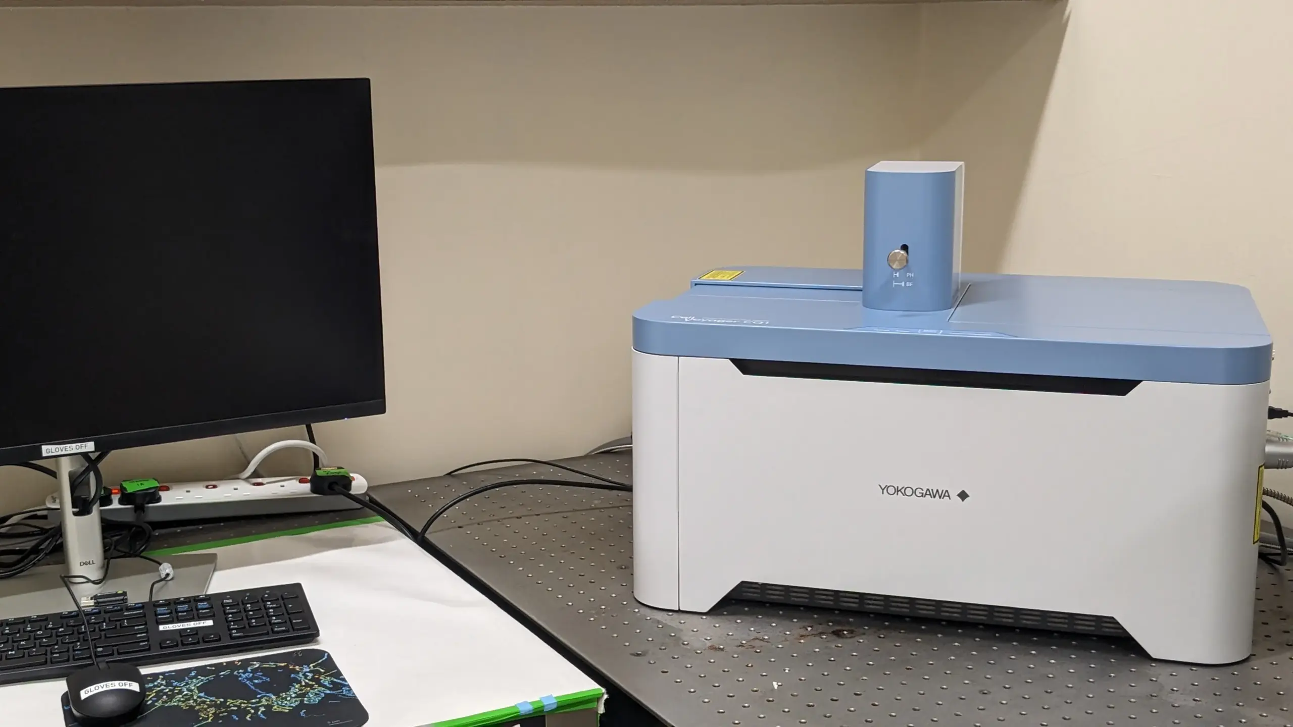
Yokogawa CQ1 Spinning Disk Confocal Microscope
Illumination: 405nm, 488nm, 561nm, 640nm
Applications:
- High-throughput 3D imaging
- Slide scans (including cover glass chambers)
- Well-plate scans: Microtiter plates or 35mm/60mm Petri dish
- Z-stack
- Live-cell imaging
- Time lapse with Multi-position capability
- High Content Analysis Software System CellPathFinder (Eg. Co-localization, Colony measurement, Spheroid Differentiation, Particle analysis, etc.)
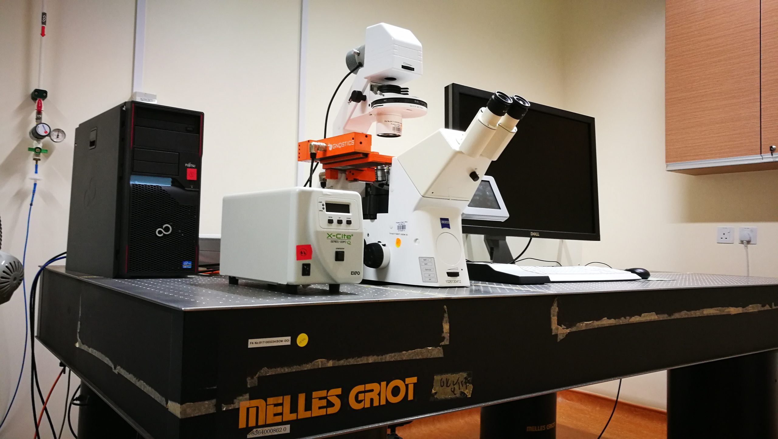
TissueGnostics: TissueFAXS Slide Scanner
Illumination: 365nm, 488nm, 545nm, 643nm, 710nm
Applications:
- High throughput bright-field and epifluorescence imaging
- Slide scans (e.g. H&E staining, Immuno-histochemistry)
- Well-plate scans
- Microarray scan
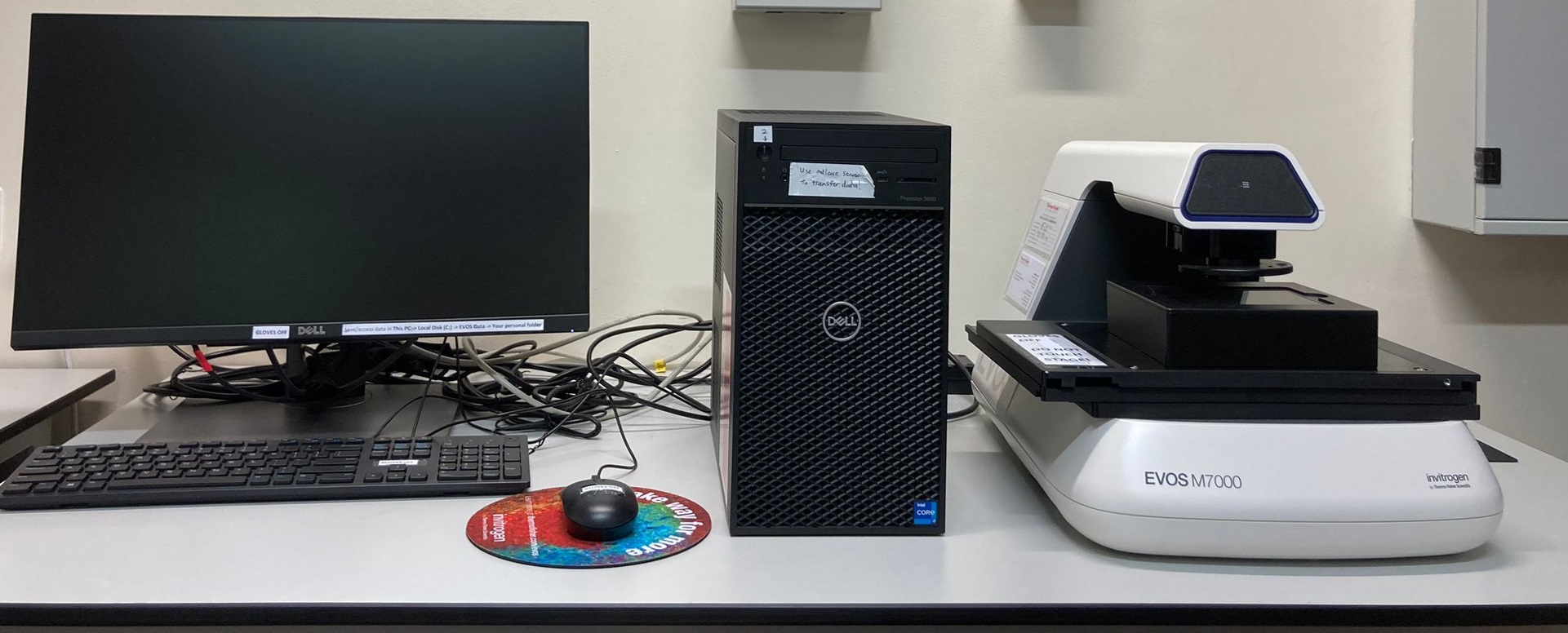
EVOS M7000 Brightfield Microscope
Light Source: LED light with DAPI, GFP, RFP, Cy5 Light Cubes
Applications:
- High throughput bright-field and epifluorescence imaging with stitching capability
- Slide scans: Digital or tissue sections eg. H&E staining, Immuno-histochemistry
- Well-plate scans: Microtiter plates or Petri dishes and plates eg. Cell culture monolayers
- Super fast live video recording (~50fps)
- Ease of use
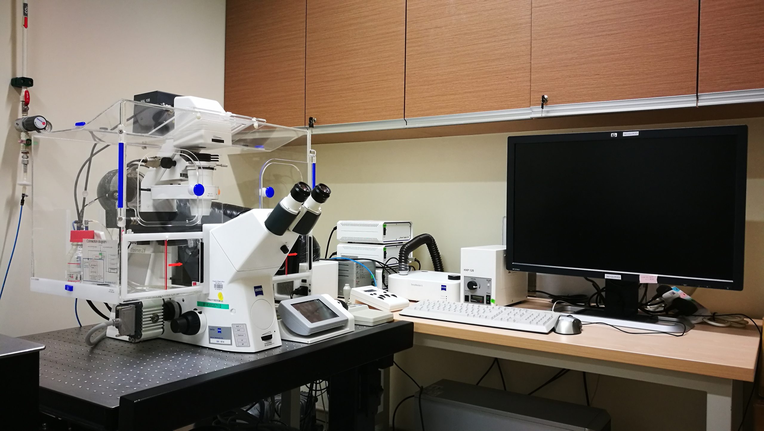
AxioObserver Z1
Applications:
- High resolution phase-contrast, DIC and epifluorescence imaging
- Time lapse with Multi-position capability
- Imaging and analysis of live cells or fixed specimens
