Over
3,000
registered users in
December 2022
Pathweb has been
accessed by users
from over
120
countries
700
virtual specimens
have been
photographed and
digitalised

Issue 46
May 2023
IN VIVO

Since its inception in 2017, Pathweb, the online pathology teaching resource, has been a go-to for students at NUS Yong Loo Lin School of Medicine (NUS Medicine), and more recently, the international medical community. What began as a labour of love for Associate Professor Nga Min En from the Department of Pathology when she built the resource has become a long-term project. It is one that has also found fans with colleagues and students, and is also shaping the way Pathology is taught and learnt over the years.
From 130 registered users in December 2018 to over 3,000 in December 2022, Pathweb has attracted users from more than 120 countries.
But Pathweb’s story harks back to 2013, when a research assistant from the Department of Radiology approached Assoc Prof Nga with an idea to photograph pathology specimens from multiple angles, to create magnifiable, rotatable virtual specimens.
Armed with a full clinical caseload but lacking experience in online content creation, Assoc Prof Nga nevertheless saw value in the proposal. She obtained a grant from NUS Medicine, and in 2014, created the first batch of virtual specimens.
Many hands make light work—the tireless team behind Pathweb
After successive rounds of grants and prototypes with resoundingly positive student feedback, Assoc Prof Nga and her colleagues decided it was time to scale up the project.
With the technical expertise of Mr Muhammed Aidil, a former staff member in Pathology, and as per his suggestion, the name and its digital avenue, Pathweb, was born in August 2017. Much of the initial website content uploads were managed by Ms Norliana Binti Abdul Aziz, the admin stalwart and the glue that holds Pathweb together.
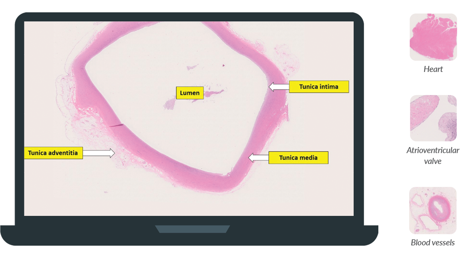
Left: The normal histology of the aorta with full annotations.
Right: Normal histology of the Cardiovascular system on Pathweb.
Aorta (Low power): The aorta is the largest artery of the circulatory system. Being an elastic artery, it has a prominent tunica media layer rich in elastic fibres and smooth muscle cells surrounding a large lumen. These features allow the aorta to expand and recoil readily in systole and diastole respectively, maintaining blood pressure and enabling pulsatile blood flow.
From a virtual pathology museum housing the fully-annotated virtual pathology pots to a plethora of educational content—mind maps, interactive quizzes and cases, a normal histology atlas and real-life cases, the immense efforts of Assoc Prof Nga and her team have turned Pathweb into an online study tool that has accumulated a large user base across the medical community, both in and out of Singapore today. But that’s not all—the team continues to work tirelessly to fine-tune and expand the content to tailor it to students’ needs.
For instance, to increase their exposure to different forms of pathology in a single disease, Assoc Prof Nga and her team photographed and digitalised more virtual specimens, from 260 to over 700 today, for students to appreciate a broader range of examples. This also comes with reconditioning degraded physical specimens, a tedious but necessary task, which requires dedicated staff who are able to perform the physically demanding tasks of reconditioning these precious specimens.
To the team’s immense delight, students enjoyed the interactive learning that Pathweb offered. “Compared to a single flat image, a 3D impression of the pots is a fantastic reproduction of what you see in person. Being better able to see the angles, textures and depths of the specimen makes the study of pathology more tangible and vivid, helping us appreciate the effects of the conditions in a way we could never do previously,” commented Tang Zhichen, a Phase II medical student.
There is no doubt that since Pathweb’s inception, benefits to the learning of pathology have been bountiful. But one student took on the challenging task of improving it, conceptualising new features that would benefit the subsequent batches of students.
Dr Darren Chua, now a first year Pathology resident, started working on Pathweb when he was in his second year of medical studies. Reflecting on his learning, he worked on ideas that he believed could value-add to his fellow students.
Veering away from pure rote learning and memorisation, he introduced the idea of Interactive Cases. With real-time self-tests with immediate feedback placed at certain points in an unravelling diagnostic scenario, students simulate the clinical work of a practicing doctor, understanding the relevance of pathology to real patients.
Another feature that Dr Chua helped to start was the Normal Histology section of the website.
“Many students find learning normal histology difficult—it often looks like abstract art to the untrained eye. Pathology, however, deals with abnormal histology, and for students who are less familiar with normal histology in the first place, recognising the abnormalities is very challenging. Hence, with this, students can have a readily accessible reference when looking at abnormal histology in various pathological conditions,” said Dr Darren Chua, first year Pathology resident who started working on Pathweb when he was in his second year of medical studies.
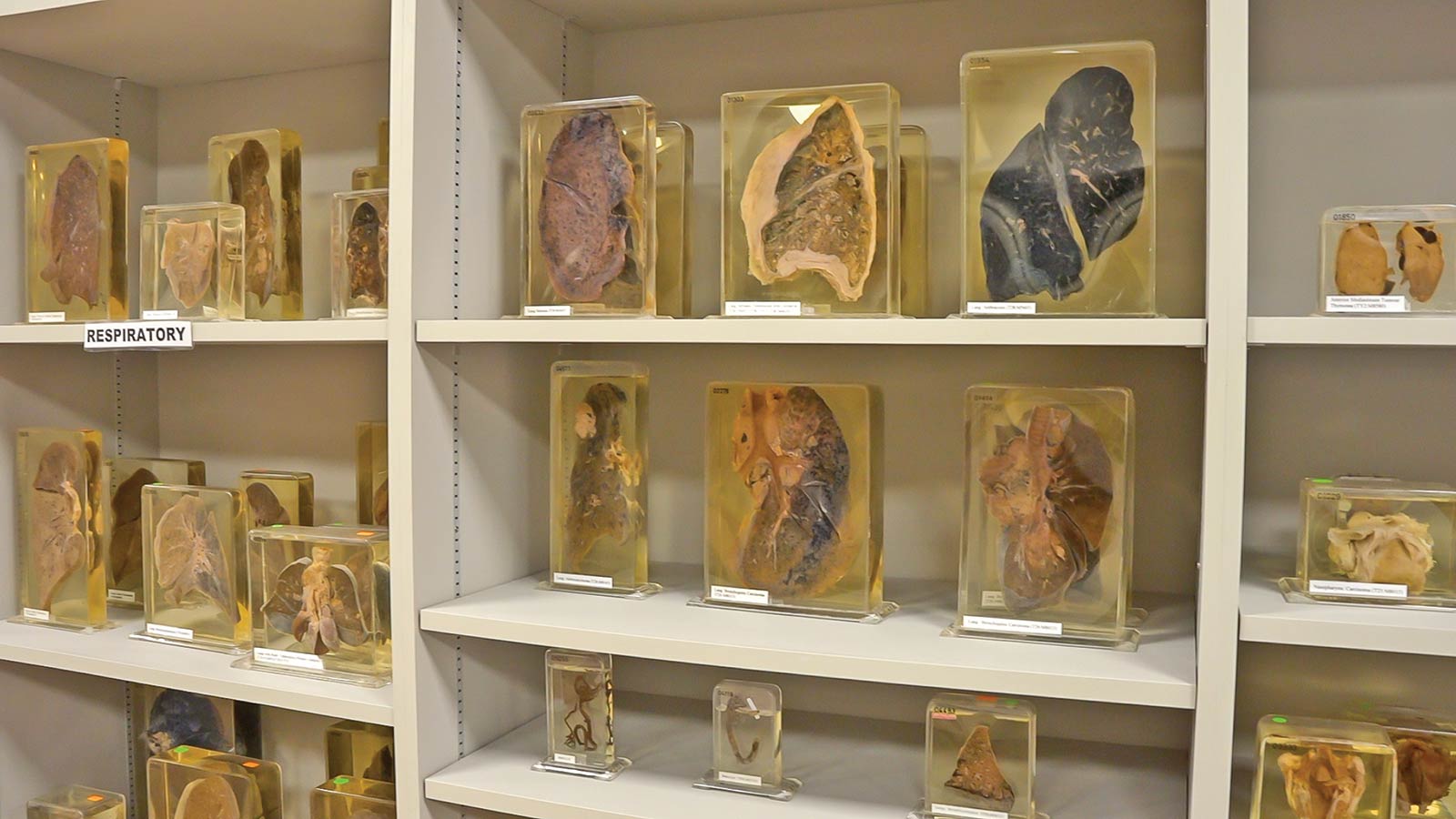
Specimens which are digitised and uploaded onto Pathweb.
The pathologist as medical detective
As doctors, pathologists are involved in the care of patients. However, this may not be immediately apparent to undergraduate students.
Through the Pathology in Action section on Pathweb, Assoc Prof Nga shares more about the daily diagnostic work of pathologists, how they handle specimens and work through cases. One might say it is clinical sleuthing. The Interactive Cases also bring out this aspect of working through real life cases and clinching the diagnosis.
“I enjoy exploring the Interactive Cases on Pathweb a lot. I recall this case about a patient presenting with a neck lump, and the interactive case guided us through taking a thorough history, making possible diagnoses, using pathological investigations and the clinical picture to come up with differentials,” said Yau Chun En, a Phase II NUS Medicine student.
“That really helped to bring pathology to life, because I could see how microscopic histological features can mean different prognoses and treatment options for the patients,” he added.
“The eye can’t see what the mind doesn’t know”
Pathweb’s popularity is explained by users’ appreciation of the online learning tool. Students enjoy the comprehensive and organised content, which is structured to accommodate different learning paces.
“I’m not that much of a fan of didactic teaching, nor do I retain much information taught in such a setting. As I explore Pathweb, I get to pause, take stock of my learning, and continue building on my existing knowledge,” said Chun En.
The study of Pathology requires high levels of attention to detail, as pathologists observe minute cells and tissues. This makes it potentially trying for students who have trouble consolidating information on the disease entity in the first place.
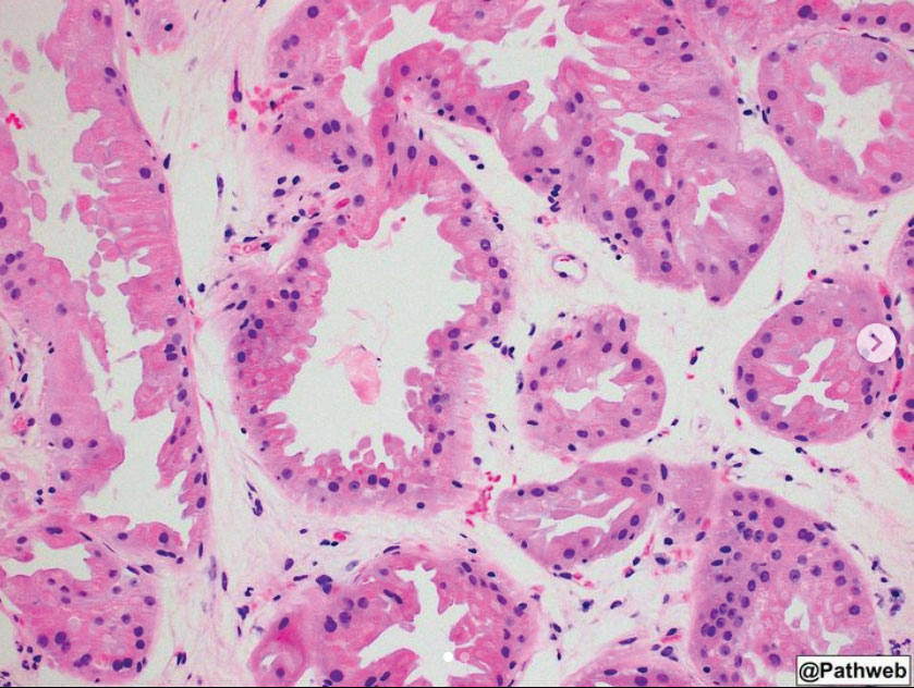
Microscopic images uploaded onto Pathweb.
For Chua Yang Jie and Wong Ting Hong, both Phase II students, the annotated specimens highlight and sensitise them to details that may be easily overlooked.
“One of our favourites on the website are the Talking Pots (concise videos on the salient features of pathology specimens preserved in containers of formaldehyde). ‘The eye cannot see what the mind doesn’t know’, so it’s very helpful to have a guided voiceover from Assoc Prof Nga as we examine the pot. It has made us much more sensitive to the subtle details which hint at the underlying disease.”
While the resources on Pathweb indeed complements in-person lessons, Assoc Prof Nga is careful that it does not become a crutch for students during lessons.
“Students do refer to the pathology virtual specimens in tutorials while I’m going through the specimens. But I don’t encourage it, I believe in the fun and mystery of figuring things out from basic principles they have learnt, so I actually instruct students to close Pathweb during my tutorials!” explained Assoc Prof Nga.
One of our favourites on the website are the Talking Pots (concise videos on the salient features of pathology specimens preserved in containers of formaldehyde). ‘The eye cannot see what the mind doesn’t know’, so it’s very helpful to have a guided voiceover from Assoc Prof Nga as we examine the pot. It has made us much more sensitive to the subtle details which hint at the underlying disease.”
An international resource, now also on social media
As a free, accessible, and comprehensive learning resource, Pathweb has attracted users from more than 120 countries.
Overseas faculty have shared that Pathweb has been a valuable resource to help them demonstrate real examples of disease to students, especially since not all medical schools have ready access to actual pathology specimens or physical pathology museums.
One overseas faculty that uses Pathweb is Associate Professor Than Than Hwte from the Faculty of Medicine, Universiti Kuala Lumpur, Royal College of Medicine, Perak.
“A huge thank you to the Pathweb team at NUS Medicine Department of Pathology, for allowing my Year 1 and 2 MBBS students and I to use Pathweb as a free resource for learning. It has really helped as a teaching aid, with clear and understandable case-based studies in Pathology,” said Assoc Prof Hwte.
“Creating and maintaining specimens can be quite costly, and specimens may not be accessible to every medical school. I’m glad that faculty members and students could access them for their teaching and learning,” said Assoc Prof Hwte.
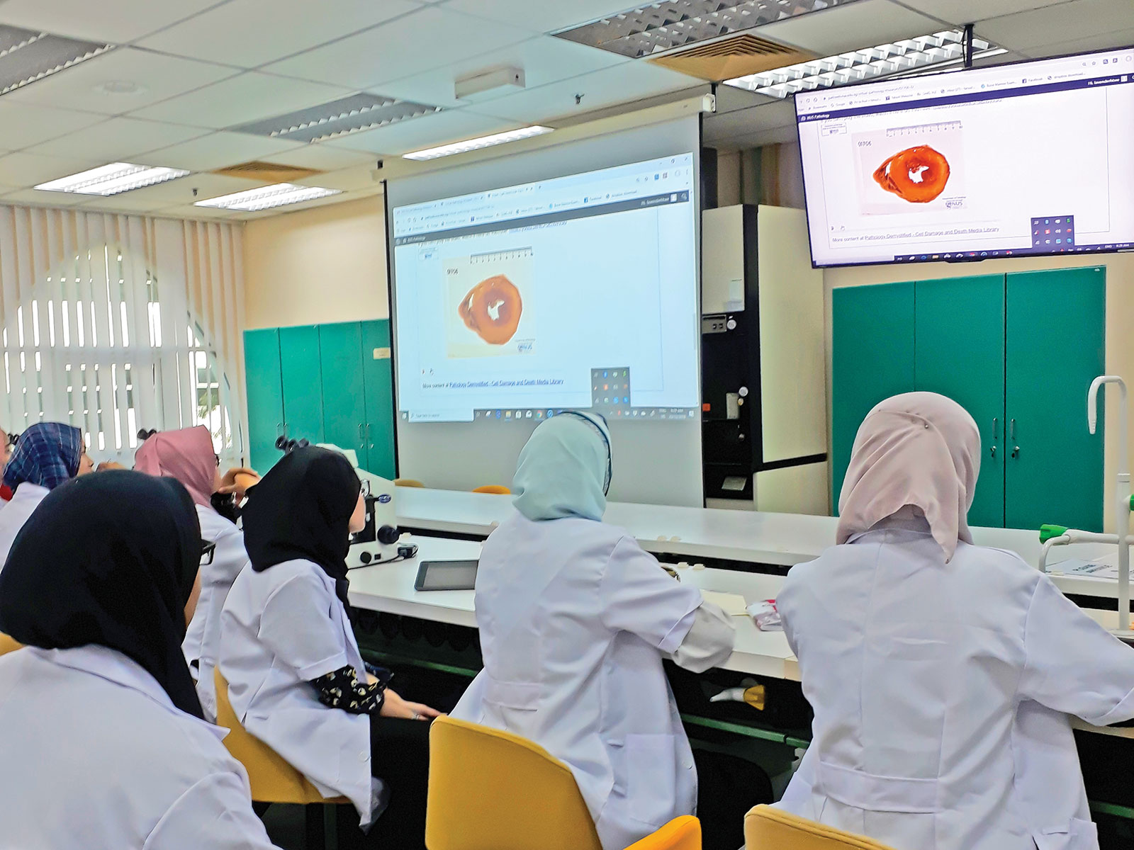
Students in Assoc Prof Hwte’s class using Pathweb in their pathology classes.
Besides the web portal, Pathweb has its own Instagram, YouTube, and Telegram channels, targeting both undergraduate and postgraduate learners in their pathology training.
Work to improve content is continuous. Tapping into Instagram, Prof Nga invited Dr Wu Bingcheng, Consultant at the Department of Pathology, National University Hospital, to populate the channel, which has over 3,000 followers today.
Creating and maintaining specimens can be quite costly, and specimens may not be accessible to every medical school. I’m glad that faculty members and students could access them for their teaching and learning.”
He employs the use of Instagram stories to post quizzes, teasing out the gaps in knowledge and difficulties that students face in real time. He also answers students’ queries on the platform, complementing their self-directed learning.
As an anatomical pathologist, he shares microscopic pictures of real-life cases from his clinical work through posts that are filled with clinical explanations.
Specialising in the head and neck region of pathology, Dr Wu explained the importance of exposing students to real life cases, since a single disease can take many forms in different organs and systems.
“The head and neck regions are anatomically critical as they contain vital structures. Within them lie several organ systems, including the upper digestive and respiratory systems, thus pathology in this area can cause significant repercussions. This explains why knowledge in and across various systems and organs in Pathology is imperative.”
Ting Hong agrees. “The exposure it has provided beyond the standard curriculum has helped me develop a sharper eye, and made me think more about pathology than I otherwise would have.”
Click here to find out more about Pathweb.
Click here to watch the introductory video on Pathweb.
More from this issue