
Issue 46
May 2023
A WORLD IN A GRAIN OF SAND
by Professor Nicholas R.J. Gascoigne, Immunology Translational Research Programme, and Department of Microbiology and Immunology, NUS Yong Loo Lin School of Medicine
A similar kind of feeling to Blake’s “World in a grain of sand” is seen in this quote from George Orwell: “Retaining one’s childhood love of such things as trees, fishes, butterflies and....toads, one makes a peaceful and decent future a little more probable1.”(Elsewhere in the essay, he makes the point that “the toad, unlike the skylark and the primrose, has never had much of a boost from poets”, but I digress).
I am still in Awe of Nature, and indeed, I’m still the kid who loves toads and butterflies, but I’ve spent my career trying to understand the immune system, which one might think is slightly less romantic. (It also doesn’t get much of a boost from poets). But consider how much the layperson has been exposed to the importance of the immune system in the last three tumultuous years; I’m pretty sure that everyone knows that a functioning immune system is important to protect you from infectious viruses. Most people now know what an antibody does and may even know about T and B cells too.
My interest, since a lucky break in getting a summer internship at a top T cell immunology lab, has been in T cells and how they recognise foreign material to make an immune response. What is most amazing about the T cell’s way of seeing the world is that it is stimulated by something that is just a tiny bit different from what it is used to—the self. The toad sees the movement of potential prey or predator because the photo-receptors in the creature’s eyes are saturated when nothing changes, so only the change registers. In a similar way, the T cell generally just sees self. That is enough stimulation to keep the individual cell alive, but not to do much. When the self-protein is slightly changed, perhaps by as little as a single amino acid, a few of the T cells will become activated by this difference, and the immune response will commence. Because the T cells see self-proteins, it is important to select them so that they don’t respond too strongly and give us autoimmunity. The T cells learn to tell the difference during their development in the organ that gives them their name, the thymus.
The protein that the T cells see is called the MHC (major histocompatibility complex), the human version of which is called HLA (human leucocyte antigen). Each HLA molecule carries a short fragment of protein (a “peptide”) in a special groove at one end of the protein. These peptides are made from worn out self-proteins that have been broken down, mostly for recycling. The T cell has a protein—the T cell receptor or TCR—on its cell surface, that can bind specifically to the HLA protein in such a position that it sees the peptide as well as the HLA protein. Each developing T cell in the thymus expresses a different TCR since the genes for the recognition part of the TCR come in lots of small modules that can be rearranged in billions of different ways, yet still produce a functional TCR.
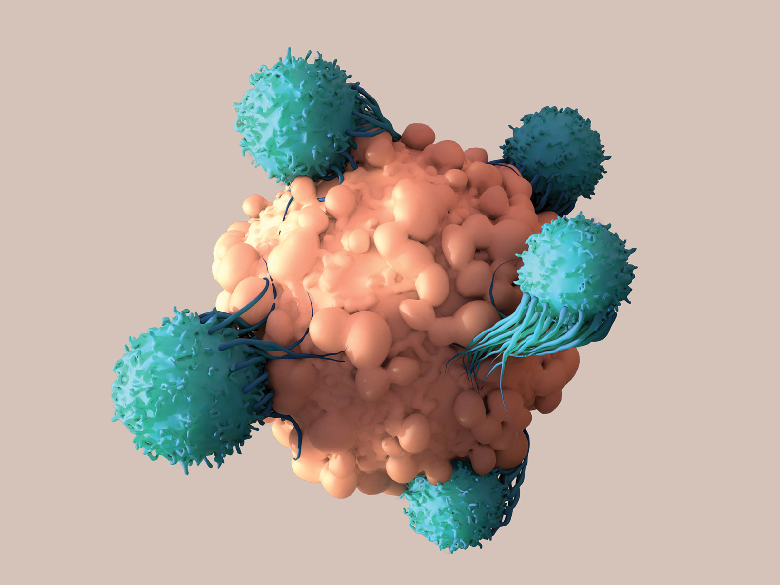
For a virus infection, there are the killer T cells, which literally kill the infected cells by squirting poisonous proteins into them, and specialized helper T cells, called TH1 cells, that make pro-inflammatory molecules (“cytokines”) that inhibit the virus’ transmission and also activate B cells to make antibodies against the virus.
The TCR of each individual cell will be exposed to the self-HLA-peptides that are expressed by the cells in the thymus. If the TCR binds too strongly to one of these HLA-peptides, the developing T cell will be over-stimulated and will die in a blaze of glory known as apoptosis or programmed cell death. If the TCR is unable to bind to any HLA-peptide it will not get any stimulus and will die “by neglect”. These two fates account for the vast majority of the cells made by the thymus, but those who bind self HLA-peptide just enough to get a little stimulation, turn on genes for survival and maturation. This Goldilocks approach means that the mature T cells that leave the thymus to go on to populate the blood, lymph nodes and spleen see self-MHC-peptide just enough to give stimulation that allows their survival.
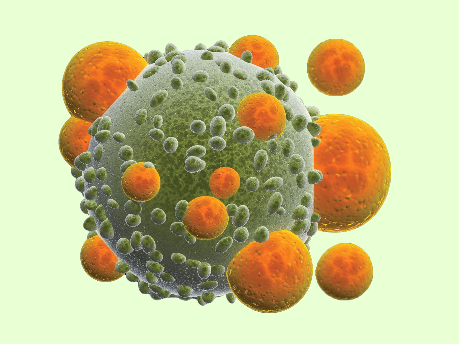
The cells can circulate through the body like this until they die or until they are lucky enough to encounter a cell that has a peptide from something different, perhaps a virus. When a virus infects a cell, it causes the cell to make viral proteins and some of these are broken down, by the same processes as the self-proteins, into peptides. Some of those peptides are incorporated into the HLA proteins, so now a TCR has the opportunity to recognise and bind to a non-self-peptide in the self-HLA. Some small number of TCRs will be able to “see” that new foreign peptide on the HLA protein, and they will get a stronger stimulus than the one from self HLA-peptide that helped keep them alive.
Now these cells turn their economies onto a war footing, they change their metabolism to a high intensity, high energy-use mode. They start proliferating, each cell dividing into millions of new cells over a period of a few days. They change from being small cells that are rather “quiet” in the sense of not really doing much, into large cells that can secrete molecules to either kill or incapacitate virus infected cells or help other cells to do so. These are “effector” T cells. They come in different forms, and the precise type of effector T cell depends on the type of infection. For a virus infection, there are the killer T cells, which literally kill the infected cells by squirting poisonous proteins into them, and specialised helper T cells, called TH1 cells, that make pro-inflammatory molecules (“cytokines”) that inhibit the virus’ transmission and also activate B cells to make antibodies against the virus.
Recently, my lab decided to find out how much time a T cell needs to see the foreign antigen in order to be stimulated, and how this changes with the strength of the interaction between the TCR and the antigen. We used a set of peptides developed from one original strong peptide and which give gradually reduced strength of T cell activation, and used T cells that all have the identical TCR. We found that the strong-binding peptide needed much less time to activate the cells to proliferate than the weaker binders, and that this was correlated with the activation of one of the molecules that turns on the metabolic changes mentioned above. We were surprised that this was not the case for the secretion of the pro-inflammatory cytokines—those didn’t need a longer time of exposure to the weaker antigens for them to be secreted.
Slight differences in how we recognise things make all the difference to how we respond. Both the T cell and the toad need to see something different to what they are used to before they can identify it as friend or foe, prey or target.
Orwell, G. (1946) Thoughts on the Common Toad. The New Republic, 20 May 1946. https://www.orwellfoundation.com/the-orwell-foundation/orwell/essays-and-other-works/some-thoughts-on-the-common-toad/ Retrieved 1 Mar 2023.
Wu, L.-z., Balyan, R., Brzostek, J., Zhao, X., and Gascoigne, N.R.J. (2023) Time required for commitment to T cell proliferation depends on TCR affinity and cytokine response. EMBO Reports 24: e54969.
More from this issue