SYNAPSE develops new technology integral to revealing brain functions
Published: 28 Sep 2023
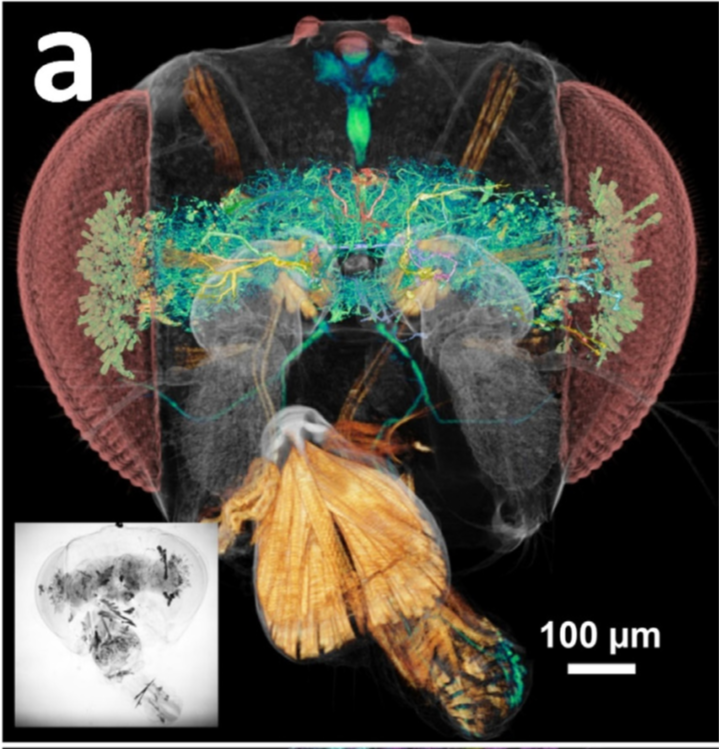
High resolution tomographically reconstructed images of neurons in the head of a Drosophila fly
Since its inauguration in 2020, the Synchrotron for Neuroscience – Asia Pacific Strategic Enterprise (SYNAPSE), a collaborative network of synchrotron facilities in the Asia Pacific region comprising more than 1,000 researchers, has been working to produce the first-of-its-kind ultra-high resolution 3D comprehensive map of the brain’s neural network.
Initiated at the Yong Loo Lin School of Medicine, National University of Singapore (NUS Medicine), the Singapore team, comprising researchers from the local scientific community, will use synchrotrons–extremely powerful x-ray sources–to trace the complex and intricate networks that cover the brain.
“Globally, brain mapping has gained impetus due to the growing impact of brain diseases. What we are setting out to do is a world-first enterprise. The images captured with unprecedented speed, clarity and granularity by SYNAPSE will form an extensive human brain map. They will show how neurons are connected and how they interact to result in cognition and intelligence,” said A/Prof Low Chian Ming from the Department of Pharmacology and Anaesthesia at NUS Medicine, who leads the SYNAPSE team in Singapore.
In its latest trope of developments, SYNAPSE has developed the Accelerated X-ray mapping Of Neurons (AXON) strategy, which enables multiscale high resolution 3-dimensional image acquisition. This technological breakthrough enables a form of nanotomography to map the neural network of the brain, as it uses x-rays to create cross-sectional images of the brain that are used to recreate a virtual model.
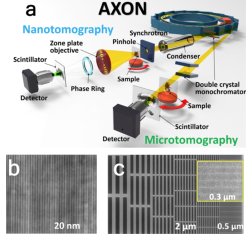
Image a: Schematic picture of how the AXON works
Images b and c: Images of test patterns whose smallest features are 20 nm (b) and 0.3 μm wide.
The AXON system is a modified (x-ray) microscope. Synchrotrons are extremely powerful x-ray sources, which are needed to produce detailed images of brain tissues, enabling us to trace the complex and intricate networks connecting the brain. The synchrotron provides the X-rays, and with the pinholes and condensers, focus and concentrate the x-rays onto the brain tissue sample. The signals that arise from the interaction between x-rays and brain tissues are selected by the zone plates (an optical device used for focusing light) and phase rings, while the scintillators convert these signals into visible light to be captured by the detector. This detector then captures the ultrafine details in brain tissues.
The capability of AXON to map whole-brain neural networks was demonstrated through the following:
- Its fast image acquisition for large volumes (less than 0.1s per projection)
- Multi-scale resolution
- 3-D resolution
- Labeling a large number of well-separated neurons
- Visualizing multiple types of internal tissues
- Validating the morphology (the form of shape of neurons) identified
- The extraction of quantitative morphology parameters for the whole brain, and statistical analysis of the results.
Significant developments include the new Brain Imaging Beamline (BIB) of the Taiwan Photon Source (TPS), which was commissioned and opened to users in 2021; and the creation of MAXWELL (Microscopy by Achromatic X-rays With Emission of Laminar Light), the brainchild of Taiwan and Japan (SPring-8)’s collaboration with Singapore. The adeptness of simultaneously capturing x-ray and visible light data from the same sample, with high resolution in 3D, represents significant advancement in the pipeline of large brain mapping.