Immunoflourescence Services
The Medical Illustration unit comprising the photomicrography unit as well as the graphics section works hand in hand with the immunohistochemistry laboratory to provide this service. Immunofluorescence microscopy has become a vital tool for the diagnosis of immunologically mediated diseases especially in kidneys, skin dermatoses, as well as other organs.
The Department of Pathology is well equipped with two epifluorescence microscopes for studying FITC antibody detections of C3 complement, IgA, IgG, IgM and other immune complex deposits in cryosections.
The microscopes are:
The Olympus BX51TRF Microscope is a transmitted light/reflected light specification model with a reflected fluorescence system. It has a HBO 100 watt mercury arc light source, and UPlan objectives: 10x, 20x, 40, 60x and 100x (oil). Up to six fluorescence mirror units can be mounted on the reflected light illuminator. Each filter unit includes a dichroic mirror, barrier filter and excitation filter that have been combined according to the excitation method. The mirror unit turret is set with: 1) Light microscopy; 2) FITC fluorescence microscopy; and 3) Ultra violet fluorescence microscopy. It has a DP 70 camera system mounted on the trinocular viewing tube for taking photomicrographs, using DP Controller and DP Manager.
The Olympus BHS Microscope is fitted with BH2-RFC reflected-light attachment, 100-watt mercury arc light source; objectives combinations are DplanApo UV - 10x, 20x, 40x, 60x (dry) and 100x (oil), and a DP 20 camera system for photomicrography. It has a full range of filter cubes for wavelength excitation-emission characteristics for major applications in fluorescence microscopy.
For examples: Ultraviolet (U) peak excitations at 334 and 365nm for:
- Observation of autofluorescence
UV immunofluorescence for general pathology, bacterial specimens, etc.
Violet (V) peak excitation at 405nm for:
- Tetracycline stain for studies of bone, teeth, etc.
Blue-violet (BV) at 405 – 435nm for:
- Thioflavine T for amyloid deposition
- Thioflavine S for lymphocyte study
- Acriflavine for nuclei acid
Blue (B) 435 – 450nm, and Interference Blue filter (IB) near 490nm for:
- FITC immunofluorescence (IB near 490nm)
- Rhodamine fluorescence
- Acridine orange stain for nuclei of cells
- Auramine stain for tubercle bacilli, etc.
- Immunofluorescence for detection of spirochetes
Screening of Immunofluorescence Preparations
Qualified laboratory officer (medical technologist) does the technical work of cutting and staining cyrosections for immunofluorescence microscopy. The officer is also trained to conduct the preliminary screening of immunofluorescence preparations to weed out the negatively stained slides. The pathologist will then interpret the sections with positive staining, and the results of the intensity and pattern of immunofluorescence are recorded. Colour photomircographs are taken for further study, publications, and clinico-pathological case discussions.
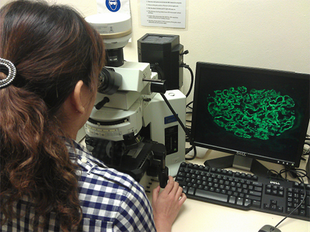
A laboratory officer conducts preliminary screening of immunofluorescence preparations. Inset: glomerulus stained with IgG, showing 3+ intensity staining in a renal biopsy of a patient with lupus nephritis (SLE).
Immunofluorescence Microscopy
1. Definition
Immunofluorescence microscopy is a laboratory technique that allows one to identify particular antigens (or antibodies) in tissues or on cells microscopically by the binding of a specific antibody that has been labeled (conjugated) with a fluorochrome.
2. Purpose
To detect the location and relative abundance of any protein for which you have an antibody. Detection of immunoglobulin and complement deposition, especially in renal and skin biopsies.
3. Principle
This entire process is the ability to visualize the antibody when a fluorescent dye that is covalently attached to the antibody is used, a light illuminates the fluorescent dye, it absorbs the light and emits a different colour light which is visible to the investigator and can be photographed through a microscope.
Fluorescence occurs when the fluorochrome absorbs the light thereby causing an electron to jump to a higher energy level (excited state). When the electron falls back to ground state, a photon is emitted. The wavelength of the emitted photon is determined by the wavelength of the absorbed photon but will always be somewhat longer due to the fact that some of the absorbed energy is dissipated in non-fluorescent ways. Fluorescein isothiocyanate (FITC) is the ones used for immunofluorescence microscopy. FITC is a green-emitting fluorochrome.
4. Phenomenon of fluorescence
Electron in the shells of fluorochrome shift shells or position when hit by excitation light (short wavelength)
When organized electrons return to their original shells position, energy gained from excitation is emitted as 2°fluroscence (longer wavelength)
5. Spectrum or white light
It covers a wavelength or about 400 cm – 750cm. Ultraviolet rays (450 – 490 um) have energy and shorter wavelength. Falls within visible spectrum, ultraviolet light f shorter wavelength is used for excitation.
6. Fluorescence Microscopy
In order to visualize the fluorescence emitted by a flurochrome, a special microscope referred to, as a "Fluorescence Microscope" is needed. This fluorescence microscope is equipped with a high-energy light source and is designed using a series of filters (exciter and barrier) that only allow light of certain wavelength to pass. The exciter filter us designed so that it filters out all excitation light except light that matched the flurochrome being used. In similar fashion, the barrier separated the weaker emitted light from the brighter excitation light. It also consists of a dichroic mirror to reflect excitation wavelength and pass omission wavelength. All these are housed in a vertical illuminator within the microscope.
7. Specimen
- Lesion or organ biopsy e.g. skin and kidney must be unfixed and delivered immediately to the laboratory
- If transport medium or saline can be used to prevent the drying of specimen if the laboratory is distant or delivery is delayed.
- On no account should they be placed in a container of formalin
- The samples must be sent to the laboratory immediately, with a completed request form indicating the need for immunofluorescence.