Hematoxylin and Eosin Stain Services
The histopathology laboratory in our department provide services of processing tissue into paraffin blocks, sectioning of tissue onto glass slides and routine hematoxylin and eosin (H&E) staining and various special staining services.
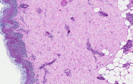
Hematoxylin and Eosin Stain (H&E)
The HE stain is a routine stain used in the histology laboratory used for studying histologic morphology in paraffin and cryosections. Hematoxylin will stain nuclei of cells blue to bluish-purple and counterstaining with Eosin will stain other cellular elements in tissues in shades of orange, pink and red.
Special Stains
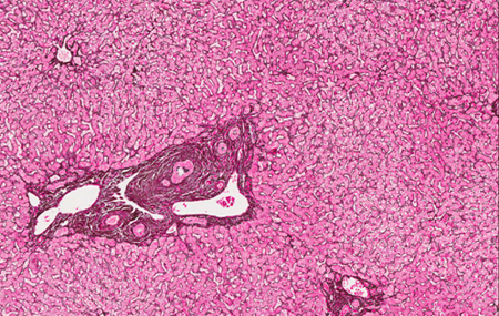
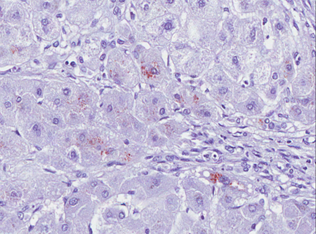
These are staining techniques using different dyes and protocols to aid in the visualization of various connective tissue, intracellular and extracellular products and microorganisms which are otherwise not easily observed under the routine HE stained slides.
Mucin staining stains
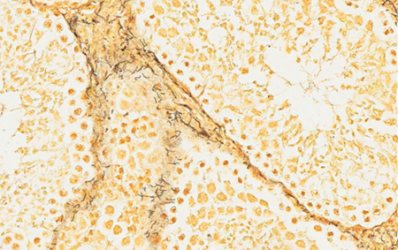
Periodic acid – Schiff (PAS) is a staining technique used to detect polysaccharides such as glycogen, and mucosubstances such as glycoproteins, glycolipids and mucins in tissues.
Southgate's Mucicarmine is a staining technique used to stain mucin secreted in various epithelial cells and connective tissues.
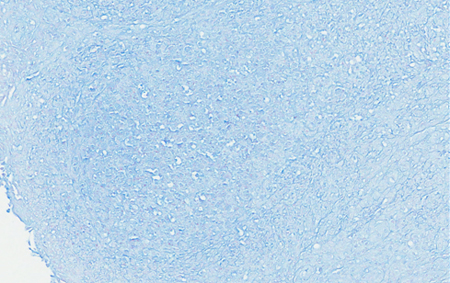
Alcian Blue is a staining technique used to stain acidic polysaccharides such as glycosaminoglycans in cartilages and other body structures.
Connective tissue stains
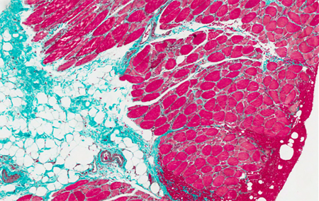
Masson trichome is a 3-colour staining technique used to distinguish various cells from surrounding connective tissue. Connective tissue is stained green, nuclei are stained dark red / purple, and cytoplasm is stained red / pink.
Gordon's and sweet method reticulin is a staining technique used to visualize reticulin fibers and used extensively in liver histopathology.
Pigments and Minerals stains
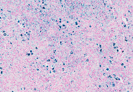
Perl's Prussian Blue is a staining techniques used to detect the presence of iron in tissue.
Fontana-Masson stain is a staining technique used to demonstrate melanin and argentaffin granules of the digestive tract.
Rhodanine stain is a staining technique to identify copper disposition in liver for the diagnosis of Wilson's disease.
Von Kossa stain is a staining technique to identify calcium deposits.
Amyloid stains
Congo Red staining is a staining technique to demonstrate amyloid deposition.
Microorganism stains
Ziehl-Neelson stain is a staining technique used to identify acid-fast organisms, mainly belonging to the Mycobacterium genus.
Warthin-Starry (WS) stain is a staining technique for detection of spirochetes and is also used to stain Helicobacter pylori. WS stains organism dark brown to black, and the background light golden brown/golden yellow.
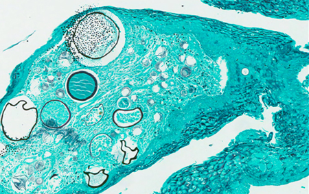
Grocott Methenamine Silver is a staining technique used widely to identify the yeast-like fungus. The cell walls of these organisms are outlined by the brown to black stain.
Giemsa is a staining technique used for histopathological diagnosis of malaria and other parasites.