Electron Microscopy
Electron Microscopy is used by the department for diagnostic work as well as for ultra structural studies of tissues, cells and their components in some areas of research. Biopsy samples of kidneys, muscle, skin, nerve, tumours and various other tissues are received, processed and prepared for Electron Microscopy within the department. Electron micrographs and viewing of the prepared specimens is done in the Electron Microscopy Unit of the Medical Faculty (NUS).

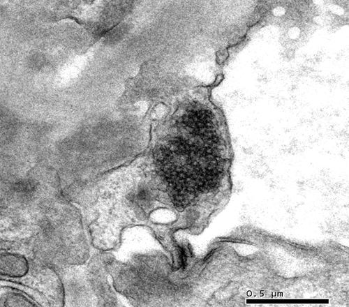
Tubuloreticular inclusions in the endothelial cell.

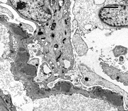
Subepithelial electron-dense deposits along the glomerular basement membrane.

