Equipment / Capabilities
TRANSMISSION ELECTRON MICROSCOPE (TEM)
FEI TECNAI SPIRIT G2 TEM
The Tecnai G2 Spirit BioTWIN operates at 100kV with a LaB6 emitter. It is an easy to use TEM designed to provide high-contrast, high-resolution imaging and analysis. It is fitted with the BioTWIN lens configurations, specifically designed to maximize contrast in images of inherently low-contrast and is well suited for exploring the 2D and 3D structures. The microscope is equipped with the FEI Eagle 4k digital camera. The bottom-mounted camera has a very large dynamic range, a high resolution and is optimized for high sensitivity.

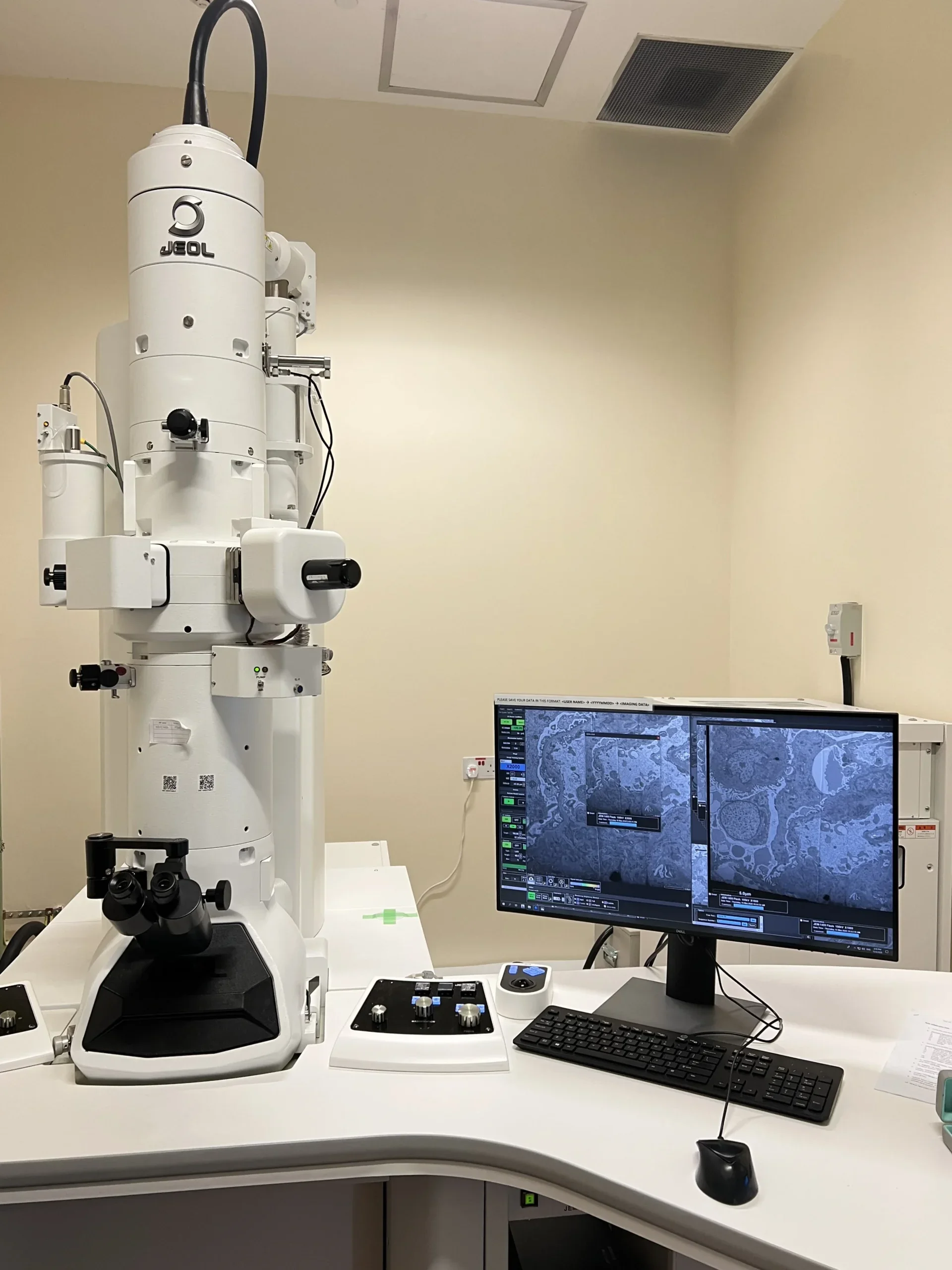
JEOL JEM-1400FLASH TEM
The JEM-1400Flash operates at 100kV with a LaB6 emitter. It is equipped with a high-sensitivity sCMOS camera, “Matataki Flash”, JEOL’s innovative high-sensitivity sCMOS camera that dramatically reduces the readout noise while possessing high frame rate. This powerful feature enables high-throughput acquisition of sharp TEM images with extremely low-noise. The Limitless Panaroma (LLP) is a montage system capable of utilizing stage drive for the field shift. This new system allows for simple capture of a montage panorama image over a limitless wide area. Thus, an ultra-wide area, high pixel-resolution image is obtainable.
THERMOFISHER TALOS L120C TEM
The Talos L120C TEM operates at 120kV with a LaB6 emitter. It is uniquely designed for performance and productivity. Talos has been widely used for different applications like 2D and 3D imaging of various samples including biological and material samples. The 4K × 4K Thermo Scientific Ceta CMOS Camera with large field of view enables live digital zooming with high sensitivity and high speed over the entire high-tension range. Talos comes with Thermo Scientific application software packages like EPU, EPU-D, E-Tomo, and Maps which allows automated data collection for different use cases and workflows, such as single particle screening, Micro-ED, tomography and large area 2D imaging.

SCANNING ELECTRON MICROSCOPES (SEM)
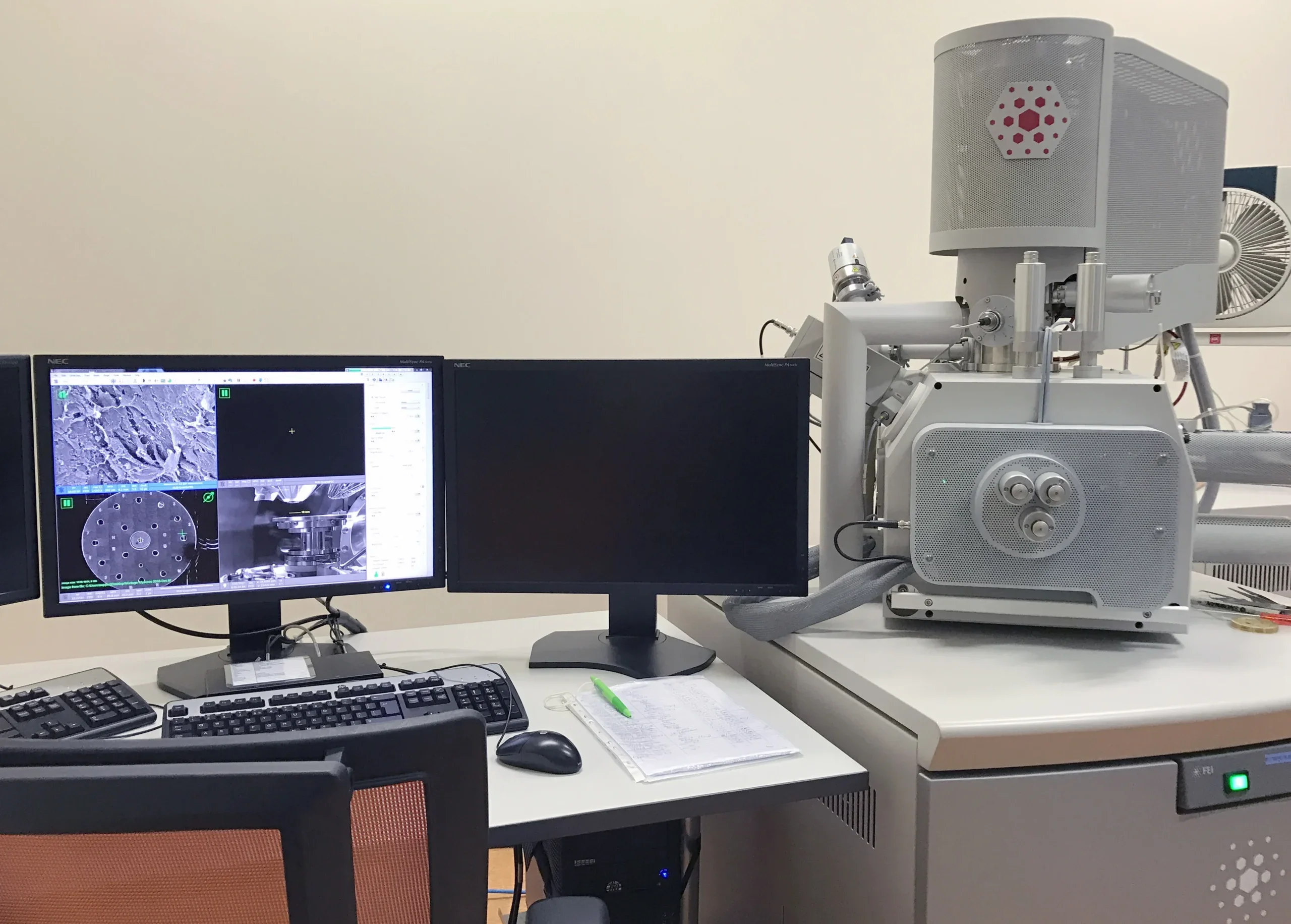
FEI QUANTA 650 FEG SEM
The Quanta 650 has a Schottky field emission gun and features a large sample chamber that enables imaging of samples of all sizes. It can be used to collect topographic information (SE mode) and observe elemental contrast (BSE mode) in the sample. Surface imaging with beam deceleration mode is possible to obtain surface and compositional information. Easy to use and intuitive controls make highly effective operation possible for novice users. The MAPS software does automatic acquisition of large overviews at any magnification, allowing high resolution imaging of large areas.
SBF-SEM: JEOL JSM-IT710HR/LV SEM WITH OXFORD EDX AND CONNECTOMX KATANA MICROTOME
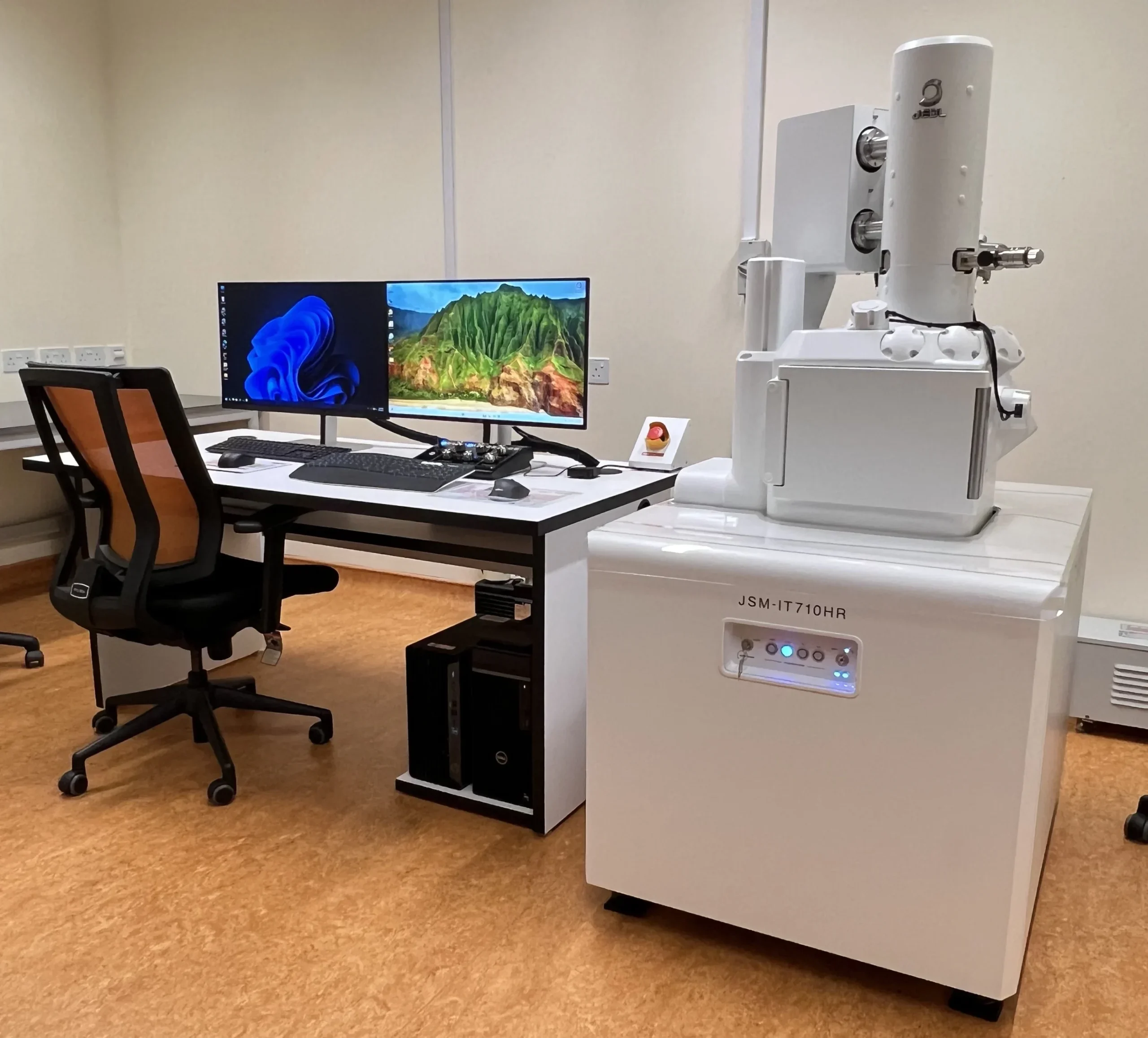
The JSM-IT710 HR SEM is a high resolution SEM with Schottky field emission gun. The simple SEM function and Zeromag mode provides the versatility to handle a wide variety of samples and workflow designed for fast results. It can be used to collect topographic information (SE and LEI mode) and observe elemental contrast (BSE mode) in the sample. The SEM is also equipped with Oxford AZtecLiveLite EDS software and Xplore 65 detector for elemental analysis.
The SEM has capabilities to perform automatic acquisition for volume EM with the Array Tomography software or Serial Block Face software, with the ConnectomX Katana microtome attached.
JEOL JSM-6701F FEG SEM
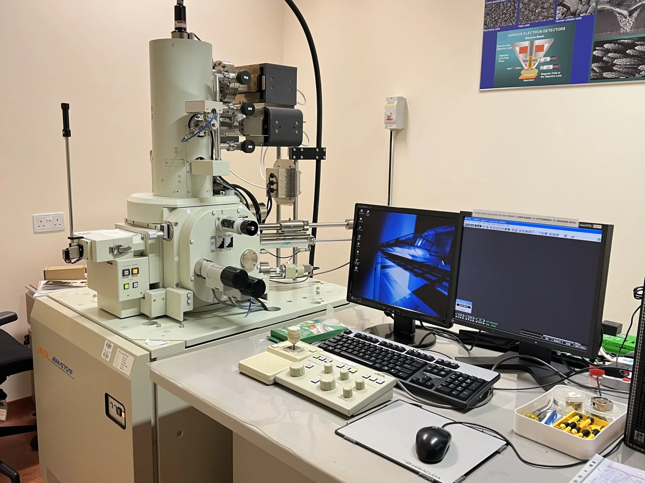
The JSM-6701F has a cold cathode field emission gun. It is suitable for observation of fine structures such as multi-layered film and nano particles fabricated by the nano technology. The high-resolution semi in-lens enables one to observe delicate specimens with minimum damage at very low accelerating voltages. At lower voltages, the fine surface structures can be observed more clearly than at higher voltages. It can be used to collect topographic information (SE and LEI mode) and observe elemental contrast (BSE mode) in the sample.
SAMPLE PREPARATION FOR MICROSCOPY
Ultramicrotomes
LEICA ULTRACUT UCT
LEICA EM UC6
LEICA EM UC7 WITH FC7 ATTACHMENT
Critical point dryers and sputter coaters
LEICA EM CPD300
LEICA EM ACE200
