A Whole New Way of Seeing the Human Body

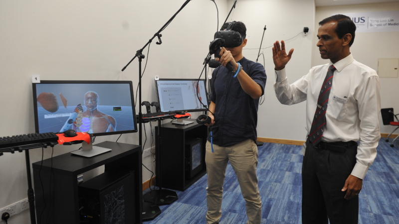
Assoc Prof Suresh Pillai giving a demonstration on how the Virtual Interactive Human Anatomy (VIHA) works.
Getting real and virtual in the study of human anatomy
The study of human anatomy is a cornerstone of medical and nursing education and at the National University of Singapore Yong Loo Lin School of Medicine (NUS Medicine), students attending classes in the subject embark on an enthralling sensory experience – they first gain hands-on experience working with human cadavers and then manipulate finely detailed, computer-generated, three-dimensional renditions of the human body and its parts in a special laboratory.
Moving from studying actual physical specimens to examining virtually simulated versions in an interactive lab, and switching again to human cadavers (dubbed Silent Mentors) allows students to gain a deeper and fuller understanding of the intricate relationships among the various body structures.
The computer-simulated human anatomy system was launched recently by the Centre for Healthcare Simulation at NUS Medicine to enhance the teaching and learning of human anatomy, said Centre director Associate Professor Suresh Pillai. Called Virtual Interactive Human Anatomy or VIHA, the system supplements and complements the traditional anatomy classes that are so essential and fundamental to medical studies. VIHA “improves the three-dimensional spatial orientation of anatomical structures as students get to interact with the virtual human body, such as removing body parts and viewing them from multiple angles. VIHA bridges the gap between textbook learning and dissection in an anatomy hall,” he explained.
In addition to prosection classes, where they study and work with cadavers that have already been dissected by experts, many students (especially those who have an interest in surgery) also attend the elective whole body dissection course. This provides opportunities for students to gain hands-on experience in cadaveric dissection.
This hands-on learning is enhanced when the students experience the cutting-edge virtual simulation technology of the VIHA system. Using Virtual Reality headsets and hand-held controllers, they are transported to a virtual dissection hall where they can perform localised or regional dissection on a virtual human cadaver to reveal underlying structures, layer by layer. Thanks to the system’s software, students are able to manipulate and mobilise joints and muscles, peel back layers of skin and tissue and peer into deeper structures like organs, blood vessels, nerves and bones. Each move can be reversed and repeated until students gain a good grasp of the relationships among the various body structures – an achievement that is not possible with a real cadaver.
“VIHA allows students to navigate the human anatomy at their own pace, reviewing and reinforcing complex spatial relationships of anatomical structures like muscles, bones,
nerves, arteries, veins and organs. Animation of joint movements has also been incorporated to highlight muscle actions in producing certain movements. This helps students with visualisation and gives them a better understanding of the connection between the various structures,” said Assoc Prof Pillai, Director of the Centre for Healthcare Simulation at NUS Medicine and also Senior Consultant at the Department of Emergency Medicine, National University Hospital.
The combination of hands-on training and virtual reality experience takes the teaching of Human Anatomy to a whole new level. Said Associate Professor S.T. Dheen, Head of Anatomy, “Traditionally, human anatomy is taught through cadaveric dissection and use of anatomical models and prosected specimens in medical education. In addition to prosection classes, where students study and work with cadavers that have already been dissected by experts, many students (especially those who have an interest in surgery) also attend the elective whole body dissection course. This provides opportunities for students to gain hands-on experience in cadaveric dissection. Although there is no substitute for cadaveric dissection in learning human anatomy, incorporating recent technological advancements like VIHA in pedagogy certainly enhances the learning process and transforms the learning environment. NUS Medicine offers the best of both worlds to Gen Z students who are generally tech savvy!”
Phase I Medicine student, Sheikh Izzat B Z-A Bahajjaj, agreed. “I find the VIHA experience very effective and practical in my learning of anatomy as it is easy to use and provides a clear, 3D visual representation of anatomical structures that are hard to visualise in the textbook. Furthermore, VIHA separates itself from the Anatomy Hall as it allows the user to freely manipulate and isolate various structures which may be difficult to appreciate, such as the courses of arteries, veins and nerves, which are difficult to observe and appreciate when using a specimen in the anatomy hall,” he said.
“While first and second year Medicine students are the first to be put through VIHA training in addition to the standard, traditional anatomy classes, the system will be progressively introduced to Phase III, IV and V medical students. This will feature more advanced features including more interactive animation, clinical pathology and self-directed questions,” added Assoc Prof Pillai. There are also plans to extend VIHA to Nursing and postgraduate students, and potentially surgical residents for pre-operative surgery planning and rehearsal of procedures.
More complex interactive training scenarios will also be introduced in the coming months: the Centre is developing the Virtual Interactive Simulation Environment (VISE), a 3D virtual environment platform that will create life-like scenarios, such as a hospital ward emergency or a mass casualty incident. These scenarios will immerse students in challenging virtual environments, where they will learn to work as teams to manage patients, applying clinical knowledge and skills that they have learned.

A student using the Virtual Interactive Human Anatomy (VIHA).
