A Labour of Love
A medical student is scrutinising a diseased heart housed in a transparent container, or a pot as it is more commonly called, while the rest of the class crane their necks in a bid for a better view. They await their turn to inspect the pot, which makes its way around the lecture room. When the specimen is finally handed to the last student at the back, the teacher has already moved on to another topic. In a different session, the class will then learn, from a separate tutor, how these diseases look on microscopic slides.
This was how pathology was taught at the National University of Singapore (NUS) Yong Loo Lin School of Medicine in the past.
Today, the learning of pathology has been enhanced and aided through technology, thanks to the efforts of Associate Professor Nga Min En and colleagues at the Department of Pathology. Painstakingly, one specimen at a time, the team has rendered more than 700 specimens in digital format and made more than 250 of these specimens available online for medical students. It is a labour of love that begun more than six years ago. It is still going on and will end when the last and final specimen has been digitised, said Assoc Prof Nga, who is a consulting pathologist at the National University Hospital.
The benefits are greatly appreciated by students, who no longer have to wait for their turn in class to view the specimens, nor borrow them from the department to study for examinations. The digitised specimens of the diseased body parts can also be viewed alongside microscopic slide images of the same disease, which helps students better understand the morphology of diseases with more clarity. Classes do not have to be split into two groups too, since both the images of the disease and the specimens can be viewed concurrently during lessons.
“The students gave us very good feedback, because they had trouble viewing the specimens clearly in the past. Now, they are able to examine the details of specimens clearly and at leisure and this also means they are able to give their attention fully to tutorials,” Assoc Prof Nga said.
“We received positive feedback from tutors as well. Most of them found the resolution (of the digitised pots) very good.”
The pathology department is home to more than 5,000 dissected and preserved specimens of human organs and tissue. While more than 700 of these pots have been digitised since 2012, Assoc Prof Nga hopes to digitise a further 1,000 specimens by the end of 2019, with the help of her team.
The digitisation process
The work to photograph the samples and then convert them to digitised images is done by a team of non-academic staff at the Department of Pathology adept at IT and photography. They are helped by students as well as NUHS Pathology residents. The latter help to check the teaching materials for the online platform, provide ideas for improvement and make value-added contributions such as annotations, adding links or cases.
The digitisation process involves a carefully orchestrated photoshoot. Staff position the specimens on a turntable inside a light box, then photograph them with a 18-megapixel camera at multiple angles. A total of 24 photos are taken of each specimen. Assoc Prof Nga then edits each image with the Photoshop software, while another team member combines these 24 images into a single file. The end result is a clear 360-degree view of the specimen in its container. It is a laborious process that takes about 45 minutes per specimen.
“It’s a labour of love. Clinical work, teaching, research, and administrative work continue. It can be a struggle to find time and energy to do this. But there are some fantastic rewards as well,” Assoc Prof Nga said.
Moving online
Seeing the potential in going digital, Assoc Prof Nga took these digitised materials one step further. In 2015, she uploaded these materials online, enabling her students to access them for their own learning.
“I believe physical pots are wonderful. I use both (physical and virtual pots) in my classes, and they complement each other. Obviously, the digital pots give much more flexibility in terms of their use; for example, students can study them any time they want to,” she explained.
The updated web resource is called Pathweb, and has two main sections. In one section called ‘Virtual Pathology Museum’, students can find both virtual microscopy slides and pots categorised into both general pathology and systemic pathology, according to their curriculum. Another section named ‘Pathology Demystified’ features mind maps, live videos, talking slides and quizzes, all hand-written, drawn or produced by Assoc Prof Nga. These materials provide students with an approach to studying pathology, and allow them to engage in self-directed learning.

Photographing specimens for digitisation.
Creating the digitised content online was not an easy feat for Assoc Prof Nga, who picked up a lot of new skills along the way, such as using new applications on her iPad to scribble notes, and voice video clips to help her students understand pathology better.
There were moments when she wondered if the digital resources would be as useful as she had believed.
“I spent days just coming up with one mind map, and I was not sure how useful people will find it. It is always a worry. You do something and invest so much time in it, and then potentially nobody might find it useful,” she said.
But the push to go digital for medical education is necessary. In the last decade, pathology labs the world over have also been digitising their slides and Assoc Prof Nga is constantly on the lookout for better ways to teach pathology.
“I am still very old-school. I use a pen and a white board to write during my class. But I think that we should explore all the means by which students learn. Tutorial classes are interactive, but online learning is another way of study. Students learn in different ways, and digital methodologies are a very huge aspect of how they learn. So we have to jump on the bandwagon.”
International fans
Since the launch of the pathology website, Assoc Prof Nga has received a fair bit of positive feedback from students and colleagues alike. Even students and academic staff from Italy and Egypt are registering for access to the site.
“I find the work very rewarding when people tell me they find (the resources) useful. A couple of times they write me an email, or in the feedback form. When I share this at overseas pathology conferences, some faculty members come to me and say that this is something very useful, and that they would like to use it,” she said.
So should users pay? “I don’t really want to make it into something people must pay for, even though it may make sense at a different level. It is like teaching – you teach and you don’t expect to get a big paycheck out of it, you do it because you want to pass on knowledge that is helpful or that will make a difference. If these resources can play a role in helping students understand pathology better and hence become more confident practitioners in the field of medicine, then, for my part, that would have made this endeavour hugely meaningful,” she said.
Phase IV Medicine student Teo Jun Hao used the Pathology Demystified website when he was learning General and Systemic Pathology in Phase II. He also used the online resource as a supplement to his textbooks and lecture notes when he needed more guidance on the topics.
“The website was really helpful in providing me a more holistic picture of what I was learning in Pathology, through the real life cases and sections on clinical-pathological correlations provided in almost every topic there. The online quizzes were also really helpful in allowing me to learn about and correct the many misconceptions I had about Pathology!” Jun Hao said.
“I think the resources on the website are very comprehensive and are a great learning tool that all medical students should explore and utilize when learning Pathology!”

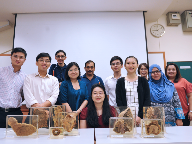
The team behind the Pathology online portal: Assoc Prof Nga Min En (seated), NUHS Pathology residents, and non-academic staff at NUS Medicine’s Department of Pathology. (Left to right) Dr Gideon Tan, Dr Nicholas Tan, Mr Muhamad Aidil, Dr Gwyneth Soon, Mr C. Rajendran, Darren Chua (NUS Medicine student, class of 2021), Dr Tan Hui Min, Ms Tan Bee Fong, and Ms Norlela Bte Mohamed, Ms Norliana Bte Abdul Aziz. Absent: Ms Tran Anh Phuong (research assistant) and Dr Noel Chia.
