Issue 44
Nov 2022

IN VIVO

By WC Wong, MBBS, PhD and Associate Professor Samuel SW Tay, Department of Anatomy, Yong Loo Lin School of Medicine

in-vivo-01-article-banner-4x3

The remote past

When the Straits and Federated Malay States Government Medical School opened its doors on 3 July 1905 in what was to be the historical beginning of medical education in the region, 16 young persons presented themselves for the full five-year course that would lead, on successful completion, to their qualification as medical practitioners1 and in the words of one of the recommendations of the Kynnersley Commission of 1902 on the system of English Education in Singapore, “supply the demand for Assistant Surgeons and General Practitioners among the native population and the poorer inhabitants”.2

As remembered 60 years later by one of the seven successful candidates who passed the final examination in Medicine, Surgery and Midwifery in the shortest prescribed time in 1910, the late Dr Chen Su Lan, “The Medical School was born without a flourish of trumpets”.3 Indeed, as measured by the subsequent evolution of the medical school and the achievements of its graduates, the original educational and professional objectives must seem to contemporary eyes relatively modest and circumscribed.

For some who yearn nostalgically for those simpler times, one fact is worth pointing out. The Government, in its reply to the signatories of the memorandum of 1904 that petitioned for the establishment of a medical school stated that the candidates for admission should “have passed the seventh standard in one or other of the secondary schools of the Colony or the Federated Malay States”.4 Standard 7 was the highest achievable attainment in English schools at that time. Primary school comprised two primary classes and Standard I, elementary school comprising Standards II to V, and secondary school comprised standards VI to VII.2 This meant that the 16 students in 1905 had an average age of 14 years (assuming that schooling began at age 6) and that of the seven successful licentiates in 1910, the average age was no more than 19 years. Viewed in our contemporary context, young men of the latter age would still be serving their National Service unless this had been deferred.

The early years

Dr RD Keith, the second principal of the School from 1909 to 1917, had written that the medical course was based largely on the programme prevailing in England, especially London and it consisted of an initial two years in the basic sciences followed by clinical clerkships in Medicine, Surgery and Midwifery during the next three years.5 The main subjects in the first year were Chemistry, Botany and Physics. The study of elementary Anatomy and Physiology begun in this year extended into the second year.

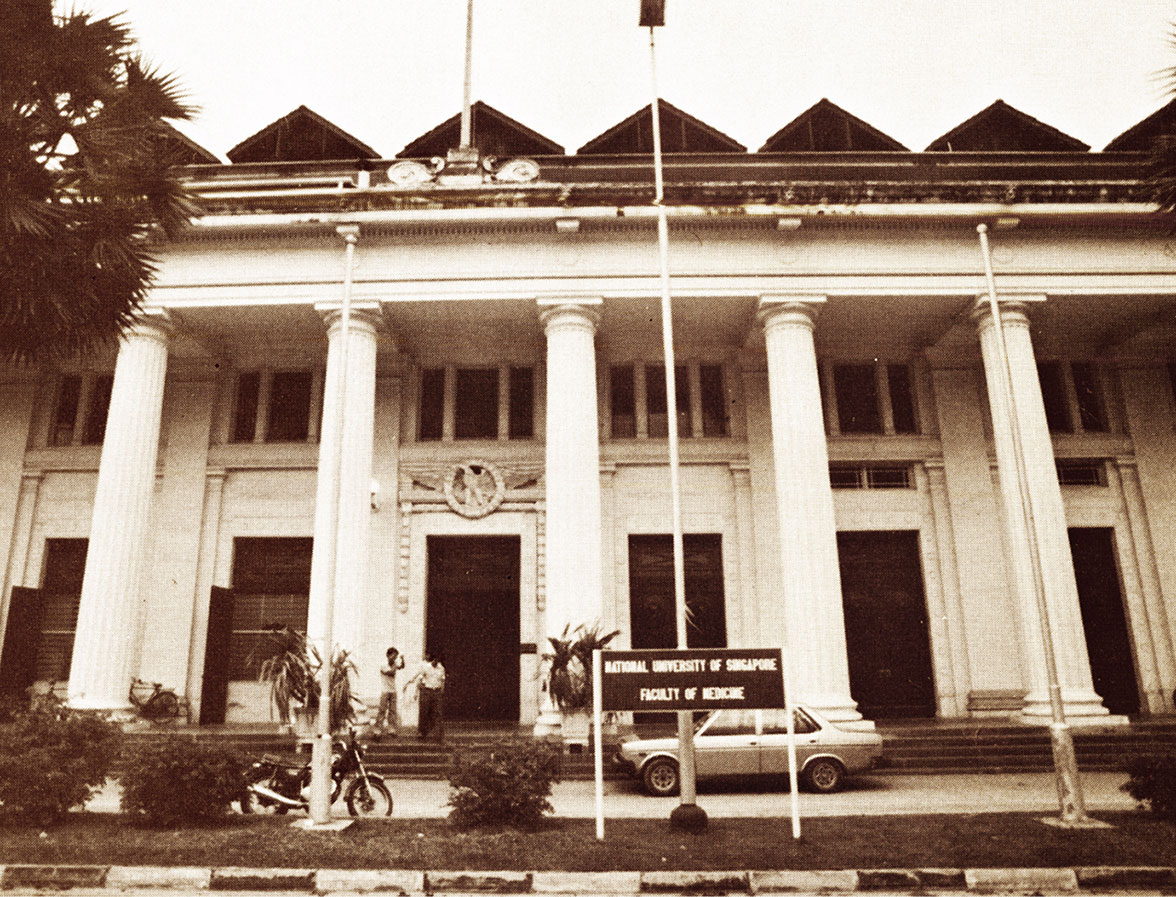
Majestic facade of the old Faculty of Medicine Building, a wonderful work of architecture to behold even today.


Dr Chen Su Lan,3 in his reminiscences, recalled that a month after the opening of the medical school, practical Anatomy began under the supervision of the first principal, Dr GD Freer. Two cadavers were available for study, a muscular young man and a young woman. How were the bodies preserved? Formalin was used as a fixative in the 1890s. In the US during the same period, various agents were used either alone or in different combinations in departments of Anatomy.6 Among these agents were carbolic acid, glycerine, arsenic, chloral hydrate, zinc chloride, mercuric bi-chloride, alcohol, potassium carbonate, sodium bicarbonate, sodium chloride, methyl spirit, formalin, potassium nitrate and boric acid. The agents were injected directly into the subjects. In some instances, the subjects were preserved by cold storage. In this connection, one of the recommendations of the committee on Medical Education in 1869 of the General Medical Council of Great Britain was that Anatomy and dissections be conducted in the first and second winter sessions of the medical course because even at that time, bodies could only be preserved in winter.7 Undoubtedly, Dr Freer and his part-time lecturers recruited from the Government Medical Service settled on a workable formula for preserving human subjects in Singapore’s perennially warm and humid conditions. Appropriately, the large building used for dissections was situated far behind the main college buildings in the refurbished former lunatic asylum.3 What were the physical conditions like? No extant description seems available. Was the building electrically lighted or were kerosene lamps still in use? Perhaps in sunny Singapore, natural light would have served. What about ventilation? Were electrically driven high ceiling fans a feature or did open windows suffice? There was nothing like the centrally air-conditioned comforts that are currently enjoyed.

Of the curriculum itself, what precisely was subsumed under “Anatomy”? Dr Chen Su Lan3 remembered that there were lectures in Osteology and that textbooks were lent to students. What were these textbooks? Of dissection guides, Professor DJ Cunningham’s Manual of Practical Anatomy had been published in 1893. Did students also have access to Henry Gray’s famous “Anatomy”? Was Histology taught under the aegis of Anatomy or was it part of the province of Physiology, as was the practice then in many medical schools in Britain? The fact that, according to Dr Chen Su Lan,3 the microscopes which arrived in 1906 were housed in the Physiology building suggested that the second possibility was probably the case. Were Neuroanatomy and descriptive Embryology also taught then? We shall have to be content with these and other questions of those early years remaining unanswered.

The recent past

As one approaches the recent past, one is on firmer ground, especially since the medical school’s observance of its 50th, 60th and 75th anniversaries. A department as such was non-existent until the first Chair of Anatomy was established in 1922.4 With this event and the provision of staff, the curriculum would have expanded to bring it in line with that of other medical schools in the British colonies, particularly those in London. By the time of the Faculty of Medicine’s diamond jubilee, the Department of Anatomy had assumed responsibility for courses for second year medical, dental, science, and third year medical as well as postgraduate (surgically aspiring) students. Besides traditional gross Anatomy, lectures and practical classes were also conducted in Neuroanatomy, Histology and descriptive Embryology.

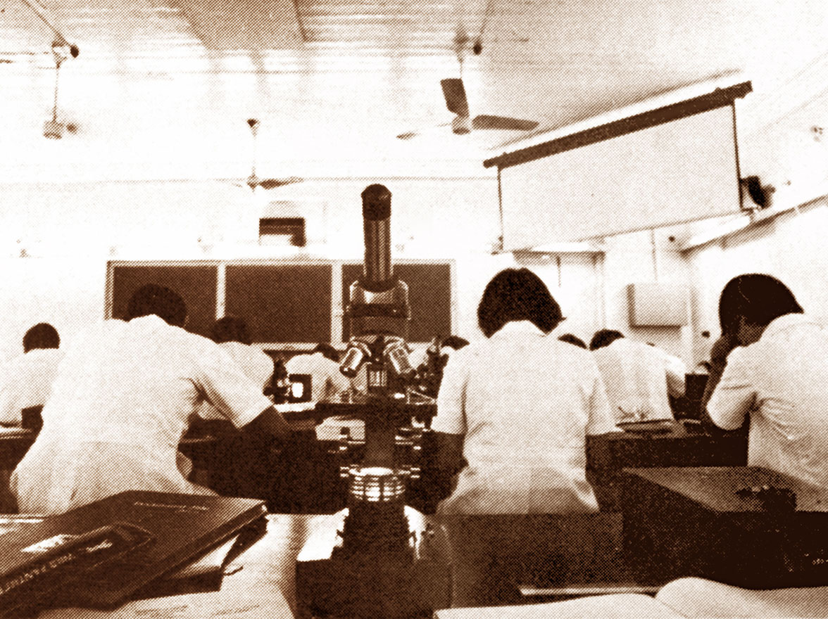
Old Histology Laboratory in the Tan Teck Guan Building.


From the mid-1960s to the mid-1990s, the course of Anatomy for medical students spanned three semesters (in 1½ academic sessions) and was conducted in such a way that gross Anatomy, Embryology, Neuroanatomy and Histology were delivered in a coordinated fashion. Emphasis was given to the clinical application where appropriate, constantly drawing the students’ attention to the professional relevance of the course. A regular feature of the course was the series of applied Anatomy lectures given by clinicians from both the public and private sectors. By the early 1980s, the gross Anatomy course was structured in such a way that at the commencement of each week, a pre-dissection lecture was given in which the week’s dissection was reviewed and the important structures that would be encountered were indicated. When the students had completed their practical study, they were divided into small groups, where individual tutors reviewed the tutorial objectives that had been made available earlier. Besides its responsibility to medical students, the Anatomy department also took part in the teaching of dental, science, pharmacy and postgraduate students.

The late 20th and early 21st centuries

In the late 1990s and early 2000s, the NUS medical curriculum was further substantially revised to meet the challenges of the time in anticipation that medical practice would be characterised by the following:

a)

The rapid expansion of biomedical knowledge.

b)

Advances in medical technology based on a better understanding of molecular and cellular processes in the human body.

c)

The rapid expansion of the Internet and related electronic media/resources.

d)

Better-educated and well-informed patients.

e)

Complex ethical issues that evolve in parallel with advances in medicine.8,9

In view of the above, a current medical student will need to have a thorough knowledge of basic medical sciences to support and strengthen future medical practice. A strong and factual foundation in Human Biology is essential for understanding human diseases. Various educational objectives were introduced recently, viz:

1)

Basic science foundation for clinical practice.

2)

Clinical competence.

3)

Communication.

4)

Appropriate attitudes.

5)

Professional development.

In line with the above educational objectives, the core M1 curriculum was organised into three tracks, namely:

1)

Structural and Cell Biology,

2)

Human Structure and Development, and

3)

Systems Biology.

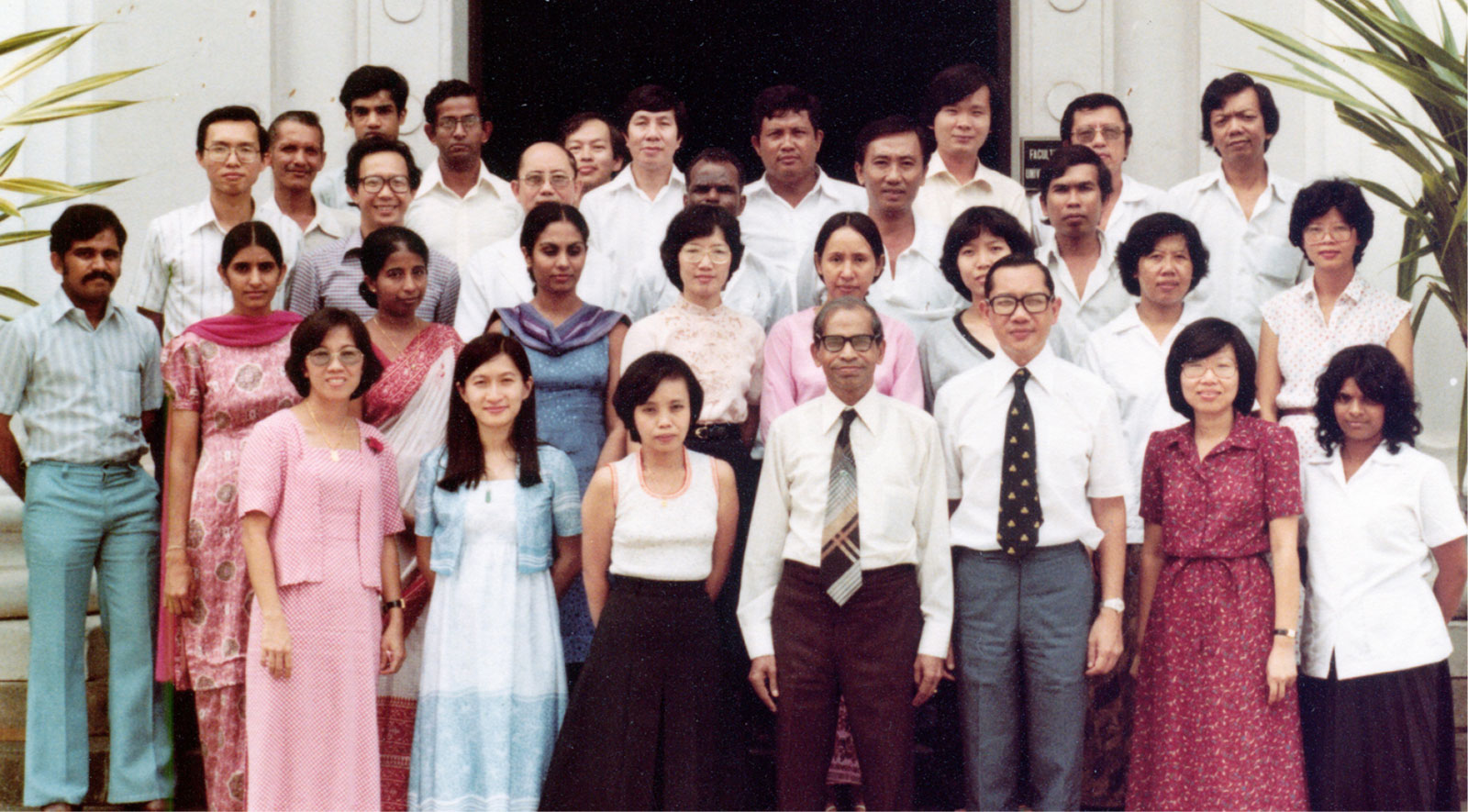
Staff members at the Sepoy Lines Campus (1980).


In this track system, Human Anatomy classically fell into the Human Structure and Development Track. The original 1½-year Anatomy programme of the mid-1990s was tailored into a one-year programme with a drastic reduction in teaching/ contact hours. The teaching of core anatomical knowledge has been further streamlined to synchronise with the teaching in Cell Biology and Systems Biology. In this way, knowledge learnt by a medical student can be integrated, with a better understanding of the structure and functions of the human body.
 

in-vivo-01-img-4

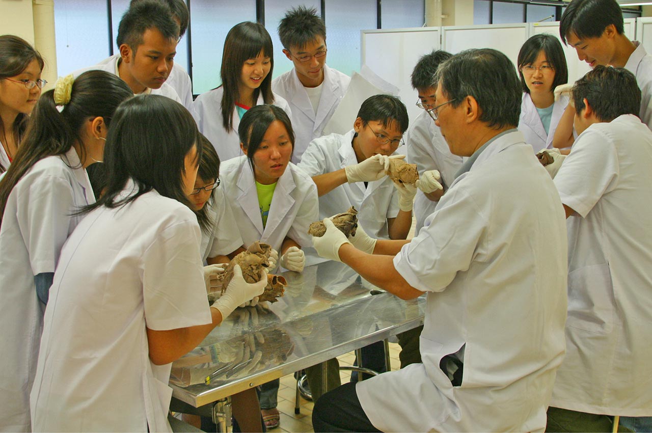
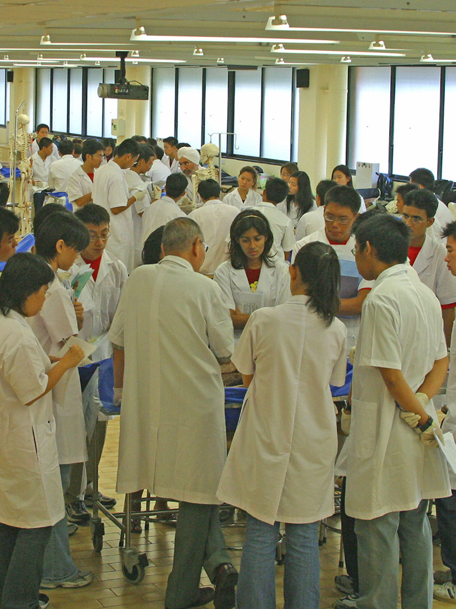
Prosection class in progress in the new Anatomy Hall.

In the Human Structure and Development Track, knowledge of gross Anatomy was imparted via dissection of the human body and prosected specimens. Students were encouraged to dissect and discover by themselves, supervised by a tutor for three hours each week, followed by a two-hour tutorial. Prior to dissection, a one-hour pre-dissection talk was given by an academic staff member from the Anatomy Department. However, a progressive fall in the availability of cadavers as well as time constraints imposed on the curriculum forced a re-examination of the teaching methodology for gross Anatomy. Recognising the human cadaver to be the most appropriate learning material, the department implemented the use of prosected cadavers in 2003/2004. Expertly prepared by trained staff and studied under the direct supervision of staff members, this method ensures the use of cadavers by students in a most efficient and effective way, using a self-exploratory approach. Besides providing a high quality of material, prosections have the advantage of allowing students to review any part of Anatomy at any time, something not possible previously with dissection, which was necessarily a destructive process.

Besides the above three tracks, medical students must attend Special Study Modules (SSMs) over a period of three weeks where they can opt to study/research an area of specialty such as Neuroscience or Toxinology in the department. In the academic year 2004/2005, the SSM was renamed the Foundation in Research Skills (FRS).

The Undergraduate Research Opportunity Programme (UROP) was initiated to attract students to have a more sustained research experience from Year 1 to Year 4. In this programme, several students have also opted to continue their research under the supervision of the staff from the Anatomy Department.

With reference to problem-based learning (PBL), a total of 10 units were introduced and later fine-tuned to the current eight units per year. Staff members from the Anatomy Department have also actively contributed to the success of PBL as facilitators and case-writers.

Staff members (past and present)

During the colonial and early post-colonial periods, the academic leadership of the department was in the hands of the expatriates, or in the contemporary phrase, foreign talents. Of the former professors and heads, one of the most fondly remembered was Professor JG Harrower (1922 to 1935), whose name has been memorialised in Harrower Hall. He has been described as an excellent teacher and research worker and he also identified himself with students and student life to a marked degree. Indeed, one of his former students, the late Professor AA Sandosham, singled him out in his memoirs for special mention as a preclinical professor who built up a strong department of Anatomy.10 Professor AR Ellis (1948 to 1960) originally from New Zealand, was a colourful lecturer, especially in his chosen special interest, descriptive Embryology. He would, in his own words, approach the subject, “from the simple to the complex”. Though not a researcher himself, he encouraged those who were interested to carry out research, particularly junior members of his staff. He did his utmost to provide the needed equipment for research, given the constraints of the time. Professor R Kanagasuntheram (1962 to 1979), originally from Ceylon (Sri Lanka) during his long tenure brought the stature and standing of the department to new heights. He was both a keen researcher and an enthusiastic teacher and endeared himself to generations of medical students, especially during his “special tutorials” held just prior to the professional examination. It was during his tenure that the first PhDs in Anatomy were instituted. Besides the three gentlemen mentioned above, attention may be drawn to others such as Dr JT Duncan and Col AJ Hull described as “brilliant teachers”,4 as well as Dr A Mohiuddin, originally from Pakistan and now settled in England.

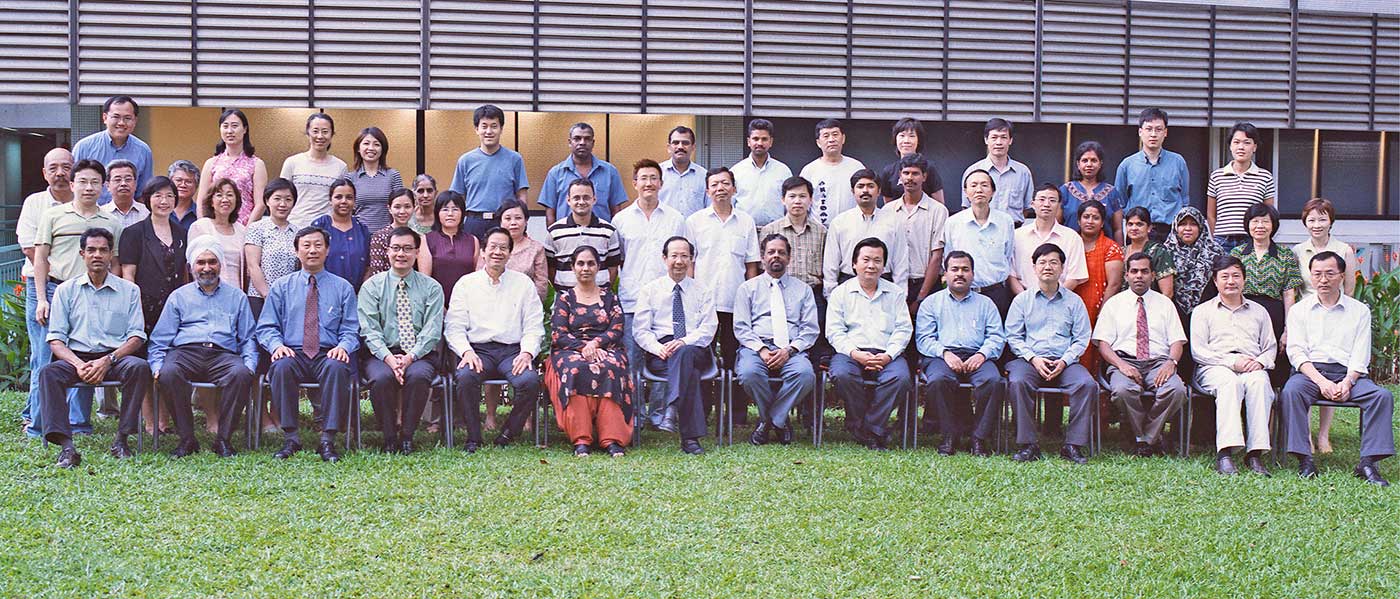
Staff members at the Kent Riadge Campus (2003).


Of the alumni of the National University of Singapore or one of its precursors—the local talents—not a few returned to make contributions of varying durations. Most of these returnees eventually pursued different careers, although some remained in Anatomy for a long time. The following list, arranged under the basic degree obtained, is nothing more than a token acknowledgement of appreciation. Licentiate in Medicine and Surgery (LMS): V Thambipillai, AWS Thevathason; Bachelor of Medicine and Bachelor of Surgery (MBBS): Chua Sui Kim, Arthur Lim Siew Ming, Lye Tong Khee, Loo Saw Kin, Wong Wai Chow, Benny Cheng Shao Lin, Quek Swee Peng, Tan Choon Kim, Sit Kwok Hung, Gurmit Singh s/o Sohan Singh, Rajendran Kanagasuntheram, Bay Boon Huat, Christopher Ang Beng Ti, George Yip Wai Cheong; Bachelor of Dental Surgery (BDS): Leong Seng Kee, Vij Sitaram, Ong Wei Yi, Ng Yee Kong; Bachelor of Science (BSc): Samuel Tay Sam Wah.

Issues in the teaching of anatomy

In a provocative essay entitled “Dilemmas in Medical Education”,11 written almost a quarter of a century ago, Professor Wong Hock Boon critiqued many aspects of medical education then, including the teaching of Anatomy. Even earlier, Dr RD Keith,5 the second principal of the medical school from 1909 to 1917, writing in 1911 on its medical course, among other comments, lamented the “old fetish” of minute descriptions of origins and insertions of muscles, remarking that inessential points should be cast aside, and more attention paid to clinical or regional anatomy, and relationships of important organs and structures. Yet, neither of these distinguished gentlemen once raised the heretical thought that Anatomy be deleted from the education of medical students and future doctors. Indeed, the answer to the question, “Why must or should Anatomy be part of the medical curriculum?” is a no- brainer. The issues in the teaching of Anatomy are not to be discovered in anatomy itself but in its what, when, how and who, i.e., what is to be taught, when should it be taught, how should it be taught and who is to teach it.

Defining how much Anatomy students should know is a perennial problem. How much is much, whether one is considering content or time allocated to teaching of Anatomy? How is a core Anatomy component for undergraduate medical education to be decided? Perhaps these questions have been misdirected in the sense that what input the teaching of Anatomy is expected to provide in the education and development of the future doctor must depend on the definition of the final product. In this regard, the Faculty of Medicine of the National University of Singapore implemented an exhaustive Curriculum Review in the academic year 1993/94.12 The five-year course has been characterised in broad outlines in which Anatomy is taught in the Human Biology Block in Year 1. More importantly, the Faculty’s stated aim is to produce doctors who “will be equipped with both scientific and clinical knowledge and skills to function effectively as house officers and have the potential to undergo further training leading to careers in primary healthcare, specialised healthcare and/or other biomedical disciplines.” Having obtained from the clinical practitioners a profile of an effective house officer, the teacher of Anatomy may then work backwards to select from the content of the subject those aspects that will contribute to this aim.

A demonstrator in action.


When should Anatomy be taught in the medical curriculum? In a significant sense, this is a non sequitur in our local university as the final model adopted is still very much an Abraham Flexner one,13 i.e., the course is divided into pre- and para-clinical and clinical blocks.

However, this is not the only approach. In his aforementioned essay,11 Professor Wong Hock Boon advocated what has come to be known in medical educational circles as vertical integration. In such a model, an organ or a system is taught in its breadth in its normal and abnormal conditions and the manifestations of symptoms and signs in disease. Professor Wong was quick to acknowledge that “such an exercise (in) integration is not easy to achieve, for if it were simple, there would have been no dilemma. Such an integration does not mean that only clinical teachers participate in teaching in the so-called ‘preclinical’ years, but ‘preclinical teachers CONTINUE teaching the students also in the clinical years during clinical teaching rounds, seminars, and conferences. We need to integrate not only student teaching, but we need also to integrate preclinical and clinical teachers. There should not be a dichotomy but there should be a cross-fertilisation.” To the best of the authors’ knowledge, there has been no attempt at such an experiment in medical education in our local medical school.


In today’s world it is unrealistic to insist that only medically qualified teachers should do so. As a general rule, those who have learnt Anatomy (wholly or partially) may teach. For those who have scant experience in Anatomy, on-the-job learning prior to taking on formal teaching responsibility will be essential.

How should Anatomy be taught? Here, the concern is whole body Anatomy as there is less controversy in the teaching of the ancillary subjects of Histology, Embryology and Neuroanatomy that come within the compass of the whole Anatomy course. In this connection, for the local medical school, as far as the teaching of Anatomy is concerned, the 2003/2004 academic year has crossed a rubicon. A sacred cow in the teaching and learning of anatomy has been slaughtered. Dissection is no more a requisite. Teachers demonstrate and students learn on prosected material. Although such an approach has been adopted in other medical schools abroad, the assessment of its success locally can only be seen in the near future. Besides the traditional cadaveric Anatomy, other aids to understanding whole body Anatomy have been and continue to be valuable such as: living Anatomy (what may be seen and felt), cross-sectional Anatomy and imaging Anatomy.14

Finally, who should teach Anatomy? In today’s world it is unrealistic to insist that only medically qualified teachers should do so. As a general rule, those who have learnt Anatomy (wholly or partially) may teach. For those who have scant experience in Anatomy, on-the-job learning prior to taking on formal teaching responsibility will be essential.

Acknowledgements

The authors are grateful to Professor Ling Eng Ang (Head, Department of Anatomy) for his support and critical comments on this article. Special thanks to Associate Professors Bay Boon Huat, K Rajendran and Dr George Yip for proofreading the manuscript. Last but not least, the authors are grateful to Mr Yick Tuck Yong for his expertise in capturing and compiling the photographs.

Click here to read more.

This article was first published in the July 2005 issue of the Annals of the Academy of Medicine, Singapore.

  • Danaraj TJ. Medical Education in Malaysia. Development and Problems. Selangor Darul Ehsan, Malaysia: Pelanduk Publications, 1988.

  • Doraisamy TR, editor. 150 Years of Education in Singapore. Singapore: Teachers’ Training College, 1969.

  • Chen SL. Reminiscences. In: Muir CS, Wong PK, editors. Sixty Years of Medical Education. Singapore: Andre Publications, 1965:31-4.

  • Sreenivasan BR, editor. History of the Medical School. In: Fifty Years of Medical Education in Malaya 1905-1955. Singapore: Craftsman Press Ltd, 1955:10-35.

  • Keith RD. A System of Medical Education. Malaya Medical Journal 1911;9:26-32.

  • Basmajian JV. The early years. In: Pauly JE, editor. The American Association of Anatomists, 1888-1987. Baltimore: Williams & Wilkins, 1987:3-13.

  • Newman C. The Evolution of Medical Education in the Nineteenth Century. London: Oxford University Press, 1957.

  • Tay SSW, Bay BH. Research Spotlight on Department of Anatomy. Singapore: Dean’s Office, Faculty of Medicine, National University of Singapore. Medviews 6 (Oct-Nov 1996):11-6.

  • New Medical Undergraduate Curriculum: Handbook for Year 1 Students, Academic Year 2000/2001. Published by Dean’s Office, Faculty of Medicine, National University of Singapore.

  • Sandosham E, Sandosham AA. The Sandoshams 1931-1981. Privately Published (Undated).

  • Wong HB. Dilemmas in Medical Education. Medical Gazette ’80, 1980:24-33.

  • Lee E, editor. A Continuity of Ideals. Commemorating 90 Years of Medical Education (1905-1995). Singapore: Armour Publishing, 1995.

  • Flexner A. Medical Education. A Comparative Study. New York, MacMillan Company, 1925.

  • McHanwell S. Anatomy education: recent advances and future developments. Symposium 25th September 2002. Anastomosis (Autumn, 2002:5-12).

More from this issue

in-vivo-01-banner-4x3