
Issue 38 / May 2021
Science of Life
When the Brain’s Housekeeping Cells Fail:
The “astrocyte” case of Jekyll and Hyde
ust like the prime minister is the most important person in a country’s governance, the neurons are the most important cells of the central nervous system (CNS). They are responsible for the transmission of information throughout our bodies and help coordinate our actions, reflexes, and sensations. But working in the background with every prime minister is a large team of secretaries, advisors, publicists, security, and more, who are vital for the minister to do his role efficiently, taking care of his schedule and every need.
Without the support of this team, the minister’s schedule would fall apart and their efficiency would be severely affected. In the CNS, the support team for the neurons are the glial cells, made up of cells like astrocytes, oligodendrocytes, and microglia, each of these cells has a specific and collaborative role. The numbers and complexity of glia evolve hand-in-hand with the complexity of the nervous system. Conversely, abnormalities in any of these cells can have a devastating impact on the performance and operation of the nervous system and this study demonstrates the interdependence of the glial cells for function.
The varied roles of glial cells
Oligodendrocytes are highly specialised cells that produce and assemble the myelin sheath, important for speedy signal conduction across the neurons, while microglia perform the role of security in the CNS, responsible for the immune response.
Astrocytes are the multitasking secretaries or housekeepers, essential for a wide range of key functions, including delivering important metabolites and energy to the neurons and maintaining a homeostatic, i.e. an optimal, balanced environment for the neuronal function. Additionally, astrocytes can become reactive in response to CNS stress due to infection, or trauma. This response can be classified into two types:
|
1) |
The A1 or cytotoxic reactive astrocytes, that usually occur in response to bacterial infections, which activate the complement immune response and cause death of other cells in the CNS. |
|
2) |
The A2 or neurotrophic reactive astrocytes, that are protective and promote repair and neuron survival. |
In other word, astrocytes seemingly have at least two personalities: the good, A2 side; and the bad, A1 side (see figure). Not surprisingly, when this multifunctional housekeeping cell has a problem, everyone has a problem—these are the cells we are interested in.
Interesting fact:
Albert Einstein had more glia
than average in his brain.
TDP-43: The ubiquitous director
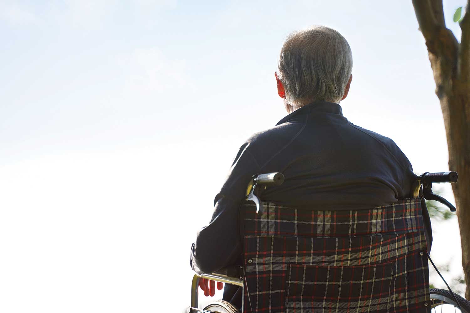
Our laboratory studies adult-onset neurodegenerative diseases, in particular amyotrophic lateral sclerosis (ALS). ALS is a progressive neuronal disease that results in a gradual deterioration and eventually death of motor neurons (the cells that control voluntary muscle movement). As these cells die, the muscle tissue wastes away while the person’s mental faculties and sensory functions remain healthy. The career of Yankee baseball player, Lou Gehrig, was cut short due to ALS. Stephen Hawking was confined to a wheelchair because of ALS.
Unfortunately, there is no effective treatment for ALS at present. How do we study this horrifying disease? The surviving motor neurons and their surrounding glia, including astrocytes, have abnormal protein aggregates containing a protein called TDP-43. Therefore, we and others have hypothesised that dysfunctional TDP-43 in neurons and glia may hold the key to understand ALS. The majority of ALS patients have been found to have mutations in TDP-43, which causes the now defective proteins to aggregate in the cell cytoplasm. These aggregates have been found not only in the neurons but also the glial cells of the patients, suggesting that the loss of TDP-43 function in glial cells also contributes to ALS disease pathogenesis.
To understand how glial cells and specifically astrocytes contribute to ALS pathogenesis, we studied the effects of TDP-43 loss in astrocytes on the CNS using mice models. Using a genetic trick, known as a the Cre-loxP system, we selectively eliminate TDP-43 from astrocytes, but not other cell types, in mice. Although the mice were born and developed normally, they displayed deficits in their motor functions, with poor balance, low grip strength and reduced mobility, accompanied by minor weight loss.
However, surprisingly, these motor deficits were not accompanied by any change in astrocyte or neuron number, no change in either proliferation or cell death was detected in these two cells types. How then was a loss of TDP-43 in astrocytes causing motor deficits? Using unbiased gene profiling and bioinformatic analysis, we found that the TDP-43 deleted astrocytes turned themselves into the bad A1-like reactive astrocytes.
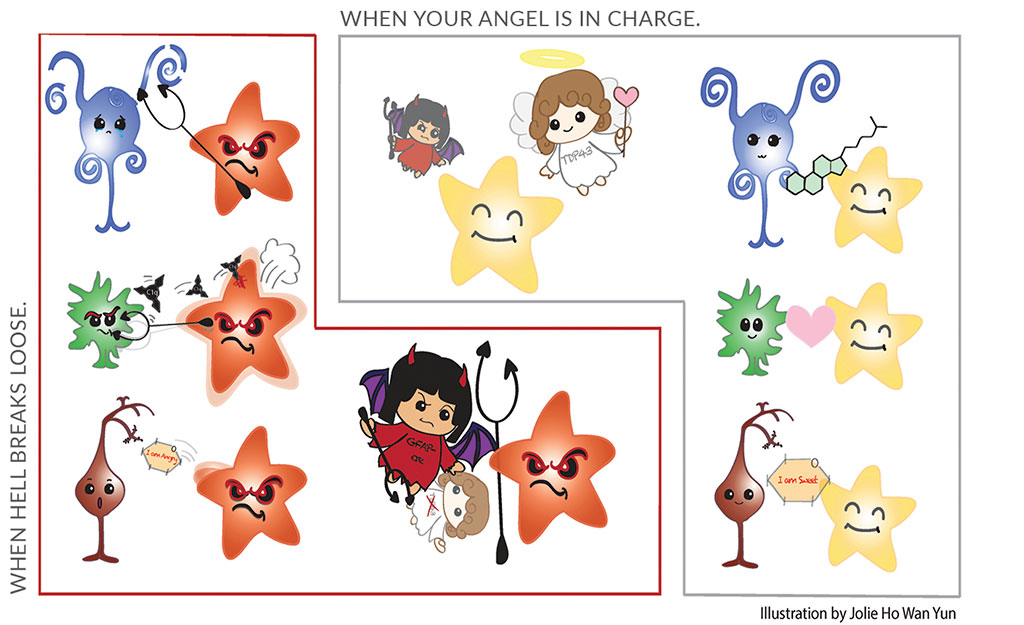
Astrocytes are “star-shaped” cells, illustrated as stars in this figure. At least two states of astrocytes are known: a good state, represented by the yellow stars; and a bad state, represented by the red stars. Astrocytes in the good state are beneficial to oligodendrocytes (represented in blue), microglia (represented in green) and neurons (represented in brown). In contrast, astrocytes in a bad state are detrimental to these three major cell types in the central nervous system. In this study, we found that TDP-43 is required to maintain astrocytes in the good state and when TDP-43 is deleted, astrocytes shift to the bad state.
The domino-effect of the missing TDP-43
Interestingly, these A1-like astrocytes were found to activate microglia (the resident security or immune cells of the CNS) resulting in an increase in expression of Complement component 1q (C1q). C1q is the initiation component of the classical complement pathway, an important immune response pathway that results in the punching of a hole in the target cell’s membrane that induces cell death. Furthermore, we found a down-regulation of the genes involved in myelin production and a reduction in the number of mature oligodendrocytes. By contrast, there were no corresponding changes in oligodendrocytes progenitor cells (OPCs). OPCs are the source of replenishing mature oligodendrocytes. The data suggests that there is a selective reduction in mature oligodendrocytes without any effect on oligodendrocyte progenitor cells.
Taken together, the data suggests that loss of TDP-43 in astrocytes causes them to activate into A1-like astrocytes that release factors which in turn activate the immune cells, microglia, to initiate the complement system. This dominos-like cascade leads to the death of oligodendrocytes in the CNS, thus reducing myelin production and hence the appearance of motor deficits in the mice. Thus, the loss of TDP-43 expression in just astrocytes led to motor deficits via a complex tri-glial dysfunction of oligodendrocytes, microglia and astrocytes (see figure).
To fall back on the support team analogy: Not only does each member of the support team need to work well individually, but every member needs to work in-sync and coordinate with each other for smooth running of the prime minister’s schedule without any hiccups. If any teammate lags behind, the performance of the entire team is affected.
Our study demonstrates that TDP-43 is required for maintaining the protective properties of astrocytes in the CNS, i.e., keeping the defensive and maintenance of astrocytes. Conversely, deleting TDP-43 in astrocytes turns astrocytes into a bad guy, as they bully microglia and oligodendrocytes. These bad interactions among astrocytes, microglia and oligodendrocytes are sufficient to cause motor deficits. Thus, one potential therapeutic strategy is to protect and promote the defensive and maintenance astrocytes.