Publications

Insights from multi-omic modeling of neurodegeneration in xeroderma pigmentosum using an induced pluripotent stem cell system

Insights from multi-omic modeling of neurodegeneration in xeroderma pigmentosum using an induced pluripotent stem cell system
Cherif Badja, Sophie Momen, Gene Ching Chiek Koh, Soraya Boushaki, Theodoros I. Roumeliotis, Zuza Kozik, Ian Jones, Vicky Bousgouni, Jo~ao M.L. Dias, Marios G. Krokidis, Jamie Young, Hongwei Chen, Ming Yang, France Docquier, Yasin Memari, Lorea Valcarcel-Zimenez, Komal Gupta, Li Ren Kong, Heather Fawcett, Florian Robert, Salome Zhao, Andrea Degasperi, Yogesh Kumar, Helen Davies, Rebecca Harris, Christian Frezza, Chryssostomos Chatgilialoglu, Robert Sarkany, Alan Lehmann, Chris Bakal, Jyoti Choudhary, Hiva Fassihi, and Serena Nik-Zainal


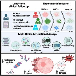
Xeroderma pigmentosum(XP) is caused by defective nucleotide excision repair of DNA damage. This results in hypersensitivity to ultraviolet light and increased skin cancer risk, as sunlight-induced photoproducts remain unrepaired. However, many XP patients also display early-onset neurodegeneration, which leads to premature death. The mechanism of neurodegeneration is unknown. Here, we investigate XP neurodegeneration using pluripotent stemcellsderivedfromXPpatients andhealthy relatives, performingfunctional multi-omicsonsamples duringneuronaldifferentiation.We showsubstantially increasedlevels of 50,8-cyclopurineand8-oxopurine in XPneuronalDNAsecondary tomarked oxidative stress. Furthermore,we find that the endoplasmic reticulum stress response is upregulated and reversal of the mutant genotype is associatedwith phenotypic rescue. Critically, XP neurons exhibit inappropriate downregulation of the protein clearance ubiquitin-proteasome system (UPS). Chemical enhancement of UPS activity in XP neuronal models improves phenotypes, albeit inadequately. Although more work is required, this study presents insights with intervention potential.

Full article: https://www.cell.com/cell-reports/fulltext/S2211-1247(24)00571-0

Insights from multi-omic modeling of neurodegeneration in xeroderma pigmentosum using an induced pluripotent stem cell system Read More »

Direct vagus nerve stimulation: A new tool to control allergic airway inflammation through α7 nicotinic acetylcholine receptor

Direct vagus nerve stimulation: A new tool to control allergic airway inflammation through α7 nicotinic acetylcholine receptor
Caroline Sévoz-Couche, Wupeng Liao, Hazel Y. C. Foo, Isabelle Bonne, Thong Beng Lu, Caris Tan Qi Hui, Wendy Yen Xian Peh, Shi-Cheng Yen, W. S. Fred Wong

Background and Purpose: Asthma is characterized by airway inflammation, mucus hypersecretion, and airway hyperresponsiveness. The use of nicotinic agents to mimic the cholinergic anti-inflammatory pathway (CAP) controls experimental asthma. Yet, the effects of vagus nerve stimulation (VNS)-induced CAP on allergic inflammation remain unknown.

Experimental Approach: BALB/c mice were sensitized and challenged with house dust mite (HDM) extract and treated with active VNS (5 Hz, 0.5 ms, 0.05–1 mA). Bronchoalveolar lavage (BAL) fluid was assessed for total and differential cell counts and cytokine levels. Lungs were examined by histopathology and electron microscopy.

Key Results: In the HDM mouse asthma model, VNS at intensities equal to or above 0.1 mA (VNS 0.1) but not sham VNS reduced BAL fluid differential cell counts and alveolar macrophages expressing α7 nicotinic receptors (α7nAChR), goblet cell hyperplasia, and collagen deposition. Besides, VNS 0.1 also abated HDM-induced elevation of type 2 cytokines IL-4 and IL-5 and was found to block the phosphorylation of transcription factor STAT6 and expression level of IRF4 in total lung lysates. Finally, VNS 0.1 abrogated methacholine-induced hyperresponsiveness in asthma mice. Prior administration of α-bungarotoxin, a specific inhibitor of α7nAChR, but not propranolol, a specific inhibitor of β2-adrenoceptors, abolished the therapeutic effects of
VNS 0.1.

Conclusion and Implications: Our data revealed the protective effects of VNS on various clinical features in allergic airway inflammation model. VNS, a clinically approved therapy for depression and epilepsy, appears to be a promising new strategy for controlling allergic asthma.

Direct vagus nerve stimulation: A new tool to control allergic airway inflammation through α7 nicotinic acetylcholine receptor Read More »

A glycolytic metabolite bypasses ‘‘two-hit’’ tumor suppression by BRCA2

A glycolytic metabolite bypasses ‘‘two-hit’’ tumor suppression by BRCA2
Li Ren Kong, Komal Gupta, Andy Jialun Wu, David Perera, Roland lvanyi-Nagy, Syed Moiz Ahmed, Tuan Zea Tan, Shawn Lu-Wen Tan, Alessandra Fuddin, Elayanambi Sundaramoorthy, Grace Shiqing Goh, Regina Tong Xin Wong, Ana S.H. Costa, Callum Oddy, Hannan Wong, C. Pawan K. Patro, Yun Suen Kho,
Xiao Zi Huang, Joan Choo, Mona Shehata, Soo Chin Lee, Boon Cher Goh, Christian Frezza, Jason J. Pitt, and Ashok R. Venkitaraman

Knudson’s “two-hit” paradigm posits that carcinogenesis requires inactivation of both copies of an autosomal tumor suppressor gene. Here, we report that the glycolytic metabolite methylglyoxal (MGO) transiently bypasses Knudson’s paradigm by inactivating the breast cancer suppressor protein BRCA2 to elicit a cancer-associated, mutational single-base substitution (SBS) signature in nonmalignant mammary cells or patient-derived organoids. Germline monoallelic BRCA2 mutations predispose to these changes. An analogous SBS signature, again without biallelic BRCA2 inactivation, accompanies MGO accumulation and DNA dam­ age in Kras-driven, Brca2-mutant murine pancreatic cancers and human breast cancers. MGO triggers BRCA2 proteolysis, temporarily disabling BRCA2’s tumor suppressive functions in DNA repair and replication, causing functional haploinsufficiency. Intermittent MGO exposure incites episodic SBS mutations without permanent BRCA2 inactivation. Thus, a metabolic mechanism wherein MGO-induced BRCA2 haploinsufficiency transiently bypasses Knudson’s two-hit requirement could link glycolysis activation by onco­ genes, metabolic disorders, or dietary challenges to mutational signatures implicated in cancer evolution.

Link to full article: https://pubmed.ncbi.nlm.nih.gov/38417710/

A glycolytic metabolite bypasses ‘‘two-hit’’ tumor suppression by BRCA2 Read More »

Blood-based biomarkers of cerebral small vessel disease

Blood-based biomarkers of cerebral small vessel disease
Liu-Yun Wu, Yuek Ling Chai, Irwin K. Cheah, Rachel S.L. Chia Saima Hilal, Thiruma V. Arumugam, Christopher P. Chen, Mitchell K.P. Lai

Age-associated cerebral small vessel disease (CSVD) represents a clinically heterogenous condition, arising from diverse microvascular mechanisms. These lead to chronic cerebrovascular dysfunction and carry a substantial risk of subsequent stroke and vascular cognitive impairment in aging populations. Owing to advances in neu-roimaging, in vivo visualization of cerebral vasculature abnormities and detection of CSVD, including lacunes, microinfarcts, microbleeds and white matter lesions, is now possible, but remains a resource-, skills- and time- intensive approach. As a result, there has been a recent proliferation of blood-based biomarker studies for CSVD aimed at developing accessible screening tools for early detection and risk stratification. However, a good understanding of the pathophysiological processes underpinning CSVD is needed to identify and assess clinically useful biomarkers. Here, we provide an overview of processes associated with CSVD pathogenesis, including endothelial injury and dysfunction, neuroinflammation, oxidative stress, perivascular neuronal damage as well as cardiovascular dysfunction. Then, we review clinical studies of the key biomolecules involved in the afore-mentioned processes. Lastly, we outline future trends and directions for CSVD biomarker discovery and clinical validation.

Link to full article: https://pubmed.ncbi.nlm.nih.gov/38417710/

Blood-based biomarkers of cerebral small vessel disease Read More »

Mechanisms of biased agonism by Gαi/o-biased stapled peptide agonists of the relaxin-3 receptor


Tharindunee Jayakody, Asuka Inoue, Srinivasaraghavan Kannan, Gaku Nakamura, Kouki Kawakami, Krishan Mendis, Thanh-Binh Nguyen, Jianguo Li, Deron R. Herr, Chandra S. Verma, Gavin S. Dawe

The neuropeptide relaxin-3 is composed of an A chain and a B chain held together by disulfide bonds, and it modu- lates functions such as anxiety and food intake by binding to and activating its cognate receptor RXFP3, mainly through the B chain. Biased ligands of RXFP3 would help to determine the molecular mechanisms underlying the

activation of G proteins and β-arrestins downstream of RXFP3 that lead to such diverse functions. We showed that the i, i+4 stapled relaxin-3 B chains, 14s18 and d(1-7)14s18, were Gαi/o-biased agonists of RXFP3. These peptides did not induce recruitment of β-arrestin1/2 to RXFP3 by GPCR kinases (GRKs), in contrast to relaxin-3, which enabled the GRK2/3-mediated recruitment of β-arrestin1/2 to RXFP3. Relaxin-3 and the previously reported peptide 4 (an i, i+4 stapled relaxin-3 B chain) did not exhibit biased signaling. The staple linker of peptide 4 and parts of both the A chain and B chain of relaxin-3 interacted with extracellular loop 3 (ECL3) of RXFP3, moving it away from the

binding pocket, suggesting that unbiased ligands promote a more open conformation of RXFP3. These findings highlight roles for the A chain and the N-terminal residues of the B chain of relaxin-3 in inducing conformational changes in RXFP3, which will help in designing selective biased ligands with improved therapeutic efficacy.

Image credit: Office of the Deputy President (Research and Technology)

Mechanisms of biased agonism by Gαi/o-biased stapled peptide agonists of the relaxin-3 receptor Read More »

Twelve tips to leverage AI for efficient and effective medical question generation: A guide for educators using Chat GPT

Twelve tips to leverage AI for efficient and effective medical question generation: A guide for educators using Chat GPT

Inthrani Raja Indran , Priya Paramanathan, Neelima Gupta and Nurulhuda Mustafa

Department of Pharmacology, National University of Singapore, Yong Loo Lin School of Medicine, Singapore, Singapore

Abstract

Background: Crafting quality assessment questions in medical education is a crucial yet time-consuming, expertise-driven undertaking that calls for innovative solutions. Large language models (LLMs), such as ChatGPT (Chat Generative Pre-Trained Transformer), present a promising yet underexplored avenue for such innovations.

Aims: This study explores the utility of ChatGPT to generate diverse, high-quality medical questions, focusing on multiple-choice questions (MCQs) as an illustrative example, to increase educator’s productivity and enable self-directed learning for students.

Description: Leveraging 12 strategies, we demonstrate how ChatGPT can be effectively used to generate assessment questions aligned with Bloom’s taxonomy and core knowledge domains while promoting best practices in assessment design.

Conclusion: Integrating LLM tools like ChatGPT into generating medical assessment questions like MCQs augments but does not replace human expertise. With continual instruction refinement, AI can produce high-standard questions. Yet, the onus of ensuring ultimate quality and accuracy remains with subject matter experts, affirming the irreplaceable value of human involvement in the artificial intelligence-driven education paradigm.

MEDICAL TEACHER
https://doi.org/10.1080/0142159X.2023.2294703

Twelve tips to leverage AI for efficient and effective medical question generation: A guide for educators using Chat GPT Read More »

SHP2 inhibitors maintain TGFβ signalling through SMURF2 inhibition

SHP2 inhibitors maintain TGFβ signalling through SMURF2 inhibition

Xianning Lai, Sarah Kit Leng Lui, Hiu Yan Lam, Yuta Adachi, Wen Jing Sim, Natali Vasilevski, Nicola J. Armstrong, Stephanie Claire Bridgeman,

Nathan Michael Main, Tuan Zea Tan, Janina E. E. Tirnitz-Parker, Jean Paul Thiery, Hiromichi Ebi, Alan Prem Kumar, and Pieter Johan Adam Eichhorn

Abstract

Despite the promising antitumor activity of SHP2 inhibitors in RAS-dependent tumours, overall responses have been limited by
their narrow therapeutic window. Like with all MAPK pathway inhibitors, this is likely the result of compensatory pathway activation
mechanisms. However, the underlying mechanisms of resistance to SHP2 inhibition remain unknown. The E3 ligase SMURF2 limits
TGFβ activity by ubiquitinating and targeting the TGFβ receptor for proteosome degradation. Using a functional RNAi screen
targeting all known phosphatases, we identify that the tyrosine phosphatase SHP2 is a critical regulator of TGFβ activity. Specifically,
SHP2 dephosphorylates two key residues on SMURF2, resulting in activation of the enzyme. Conversely, SHP2 depletion maintains
SMURF2 in an inactive state, resulting in the maintenance of TGFβ activity. Furthermore, we demonstrate that depleting SHP2 has
significant implications on TGFβ-mediated migration, senescence, and cell survival. These effects can be overcome through the use
of TGFβ-targeted therapies. Consequently, our findings provide a rationale for combining SHP2 and TGFβ inhibitors to enhance
tumour responses leading to improved patient outcomes.


npj Precision Oncology (2023)7:136 ; https://doi.org/10.1038/s41698-023-00486-6

SHP2 inhibitors maintain TGFβ signalling through SMURF2 inhibition Read More »

Isthmin-1 attenuates allergic Asthma by stimulating adiponectin expression and alveolar macrophage efferocytosis in mice

Isthmin-1 attenuates allergic Asthma by stimulating adiponectin expression and alveolar macrophage efferocytosis in mice

Jong Huat Tee, Udhaya Vijayakumar, Mahalakshmi Shanmugasundaram, Terence Y. W. Lam, Wupeng Liao, Yuansheng Yang, W. S. Fred Wong* and Ruowen Ge*

Abstract
Background Allergic asthma is a common respiratory disease that significantly impacts human health. Through in silico analysis of human lung RNASeq, we found that asthmatic lungs display lower levels of Isthmin-1 (ISM1) expression than healthy lungs. ISM1 is an endogenous anti-inflammatory protein that is highly expressed in mouse lungs and bronchial epithelial cells, playing a crucial role in maintaining lung homeostasis. However, how ISM1 influences asthma remains unclear. This study aims to investigate the potential involvement of ISM1 in allergic airway inflammation and uncover the underlying mechanisms.

Methods  We investigated the pivotal role of ISM1 in airway inflammation using an ISM1 knockout mouse line (ISM1−/−) and challenged them with house dust mite (HDM) extract to induce allergic-like airway/lung inflammation. To examine the impact of ISM1 deficiency, we analyzed the infiltration of immune cells into the lungs and cytokine levels in bronchoalveolar lavage fluid (BALF) using flow cytometry and multiplex ELISA, respectively. Furthermore, we examined the therapeutic potential of ISM1 by administering recombinant ISM1 (rISM1) via the intratracheal route to rescue the effects of ISM1 reduction in HDM-challenged mice. RNA-Seq, western blot, and fluorescence microscopy techniques were subsequently used to elucidate the underlying mechanisms.

Results  ISM1−/− mice showed a pronounced worsening of allergic airway inflammation and hyperresponsiveness upon HDM challenge. The heightened inflammation in ISM1−/− mice correlated with enhanced lung cell necroptosis, as indicated by higher pMLKL expression. Intratracheal delivery of rISM1 significantly reduced the number of eosinophils in BALF and goblet cell hyperplasia. Mechanistically, ISM1 stimulates adiponectin secretion by type 2 alveolar epithelial cells partially through the GRP78 receptor and enhances adiponectin-facilitated apoptotic cell clearance via alveolar macrophage efferocytosis. Reduced adiponectin expression under ISM1 deficiency also contributed to intensified necroptosis, prolonged inflammation, and heightened severity of airway hyperresponsiveness.

Conclusions  This study revealed for the first time that ISM1 functions to restrain airway hyperresponsiveness to HDM-triggered allergic-like airway/lung inflammation in mice, consistent with its persistent downregulation in human asthma. Direct administration of rISM1 into the airway alleviates airway inflammation and promotes immune cell clearance, likely by stimulating airway adiponectin production. These findings suggest that ISM1 has therapeutic potential for allergic asthma.

Isthmin-1 attenuates allergic Asthma by stimulating adiponectin expression and alveolar macrophage efferocytosis in mice Read More »

Multifunctional Antibacterial Nanonets Attenuate Inflammatory Responses through Selective Trapping of Endotoxins and Pro-Inflammatory Cytokines

Multifunctional Antibacterial Nanonets Attenuate Inflammatory Responses through Selective Trapping of
Endotoxins and Pro-Inflammatory Cytokines

Nhan Dai Thien Tram, Quy Thi Ngoc Tran, Jian Xu, Jeannie Ching Ting Su, Wupeng Liao, Wai Shiu Fred Wong, and Pui Lai Rachel Ee*

Extracellular lipopolysaccharide (LPS) released from bacteria cells can enter the bloodstream and cause septic complications with excessive host inflammatory responses. Target-specific strategies to inactivate inflammation mediators have largely failed to improve the prognosis of septic patients in clinical trials. By utilizing their high density of positive charges, de novo
designed peptide nanonets are shown to selectively entrap the negatively charged LPS and pro-inflammatory cytokines tumor necrosis factor-𝜶 (TNF-𝜶) and interleukin-6 (IL-6). This in turn enables the nanonets to suppress LPS-induced cytokine production by murine macrophage cell line and rescue the antimicrobial activity of the last-resort antibiotic, colistin, from LPS
binding. Using an acute lung injury model in mice, it is demonstrated that intratracheal administration of the fibrillating peptides is effective at lowering local release of TNF-𝜶 and IL-6. Together with previously shown ability to simultaneously trap and kill pathogenic bacteria, the peptide nanonets display remarkable potential as a holistic, multifunctional anti-infective, and
anti-septic biomaterial.

Multifunctional Antibacterial Nanonets Attenuate Inflammatory Responses through Selective Trapping of Endotoxins and Pro-Inflammatory Cytokines Read More »

Degradation of MK2 with natural compound andrographolide: A new modality for anti-inflammatory therapy

Degradation of MK2 with natural compound andrographolide: A new modality for anti-inflammatory therapy

Quy T.N. Tran a,b,c,1, Phyllis X.L. Gan a,2, Wupeng Liao a,d,3, Yu Keung Mok e,4, Christina L.L. Chai b,c,*,5, W.S. Fred Wong

a Department of Pharmacology, Yong Loo Lin School of Medicine, National University Health System, 117600, Singapore
b Department of Pharmacy, Faculty of Science, National University of Singapore, 117543, Singapore
c Drug Discovery and Optimization Platform (DDOP), Yong Loo Lin School of Medicine, National University Health System, 117600, Singapore
d Singapore-HUJ Alliance for Research and Enterprise (SHARE), National University of Singapore, Singapore
e Department of Biological Sciences, Faculty of Science, National University of Singapore, 117543, Singapore

A B S T R A C T

The p38MAPK-MK2 signaling axis functions as an initiator of inflammation. Targeting the p38MAPK-MK2 signaling axis represents a direct therapeutic intervention of inflammatory diseases. We described here a novel role of andrographolide (AG), a small-molecule ent-labdane natural compound, as an inhibitor of p38MAPK-MK2 axis via MK2 degradation. AG was found to bind to the activation loop of MK2, located at the interface of the p38MAPKMK2 biomolecular complex. This interaction disrupted the complex formation and predisposed MK2 to proteasome-mediated degradation. We showed that AG induced MK2 degradation in a concentration- and timedependent manner and exerted its anti-inflammatory effects by enhancing the mRNA-destabilizing activity of tristetraprolin, thereby inhibiting pro-inflammatory mediator production (e.g., TNF-α, MCP-1). Administration of AG via intratracheal (i.t.) route to mice induced MK2 downregulation in lung alveolar macrophages, but not lung tissues, and prevented macrophage activation. Our study also demonstrated that the anti-inflammatory effects achieved by AG via MK2 degradation were more durable and sustained than that achieved by the conventional MK2 kinase inhibitors (e.g., PF-3644022). Taken together, our findings illustrated a novel mode of action of AG by modulating the p38MAPK-MK2 signaling axis and would pave the way for the development of a novel class of anti-inflammatory agents targeting MK2 for degradation by harnessing the privileged scaffold of AG.

Degradation of MK2 with natural compound andrographolide: A new modality for anti-inflammatory therapy Read More »