Metabolic and inflammatory roles of glial cells in neurodegenerative and cerebrovascular diseases


Cortical cerebral microinfarcts (CMIs) are associated with cognitive dysfunction and dementia, while their evolution on sequential magnetic resonance imaging (MRI) remains unclear.
The study enrolled 490 patients (72.5 ± 7.9 years) from a memory-clinic cohort, with 5-year follow-up. Cortical CMIs were graded at baseline and year 2 to identify incident lesions and other evolutionary patterns. Cognitive function was assessed annually. Clinical events, including dementia, stroke, and mortality, were recorded with time to event.
Forty-one (8.4%) patients showed incident cortical CMIs at year 2. Additionally, 12 had CMIs becoming invisible, and six showed CMIs incorporated into new large infarctions. Baseline cortical CMIs and large cortical infarcts showed the strongest association with incident CMIs. Incident cortical CMIs were associated with cognitive decline, white matter hyperintensity progression, and incident dementia, independent of prevalent lesions.
Cortical CMI evolution may reflect dynamic changes in brain vascular pathology and represent a potential target for interventions aimed at preserving cognitive function.

Gastric cancer, the fifth most prevalent cancer globally, poses significant treatment challenges due to factors such as late diagnosis, early metastasis, limited surgical options, and the systemic toxicity of chemotherapy. Because luminal barriers are often compromised in gastric cancers , orally administered therapies that enable localized absorption and drug release represent a promising new direction for site-specific treatment with limited side effects.
We introduce a disulfide-linked thermostable exoshell system that orally delivers protein-based bioorthogonal catalytic centres directly to cancer tissues. The highly engineered exoshells effectively encapsulated and stabilized labile catalytic centres, preventing degradation in the harsh gastric environment. In vivo gastric tumors were treated using the anti-cancer properties of active metabolites of the prodrug indole-3-acetic acid (IAA) converted in situ via bioorthogonal catalysis. In vitro cell studies revealed a dose- and time-dependent inhibition of gastric cancer cell growth, irrespective of their HER2 status. This inhibition was accompanied by upregulation of mitochondrial lipid peroxidation, reduced mitochondrial membrane potential, and activation of necroptotic pathway markers such as RIP1, RIP3, and MLKL at both mRNA and protein levels. In a mouse model of gastric cancer induced by N-Methyl-N-Nitrosourea, oral administration of catalytic exoshells for 6 weeks significantly inhibited gastric inflammation and tumour polyp growth. Additionally, LC/MS/MS-based metabolomic analysis of plasma obtained from treated mice showed significant upregulation of cytotoxic metabolites of IAA. Notably, metabolites relevant to redox regulation, including alpha-tocopherol (vitamin E), glutathione (GSH), homocysteine, methyl cysteine, and cysteine sulfinic acid, were identified as the top differentially expressed metabolites, indicating potent suppression of inflammation and tumour growth. Histological analysis of gastric tissue showed a reduced number of polyps and subsequent development of gastric tumours.
Our in vitro and in vivo results demonstrated that exoshells possessed significant potential as an orally administered, titratable therapeutic platform for the management of gastrointestinal cancers.

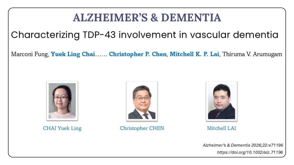
Vascular dementia (VaD) is a major therapeutic challenge. Tar DNA-binding protein 43 (TDP-43), known for its role in neurodegeneration, may contribute to VaD pathogenesis under chronic cerebral hypoperfusion (CCH). This study investigates TDP-43 dysregulation in VaD.
TDP-43 and phosphorylated TDP-43 (pTDP-43) expression and localization were assessed in a VaD animal model, neuronal cells exposed to oxygen–glucose deprivation (OGD), and post mortem human brain tissues.
Bilateral Common Carotid Artery Stenosis (BCAS)-induced CCH led to increased pTDP-43 and aberrant redistribution of both TDP-43 and pTDP-43. In vitro OGD triggered similar mislocalization. Post mortem VaD brains showed no TDP-43 abnormalities, while Alzheimer’s and mixed dementia cases exhibited marked pathology.
TDP-43 dysregulation appears early in VaD under hypoperfusive stress, distinguishing it from other dementia subtypes. These findings indicate that TDP‑43 may warrant further investigation as a potential early molecular feature of VaD.
Full Article:Â https://alz-journals.onlinelibrary.wiley.com/doi/10.1002/alz.71196
Characterizing TDP-43 involvement in vascular dementia Read More »

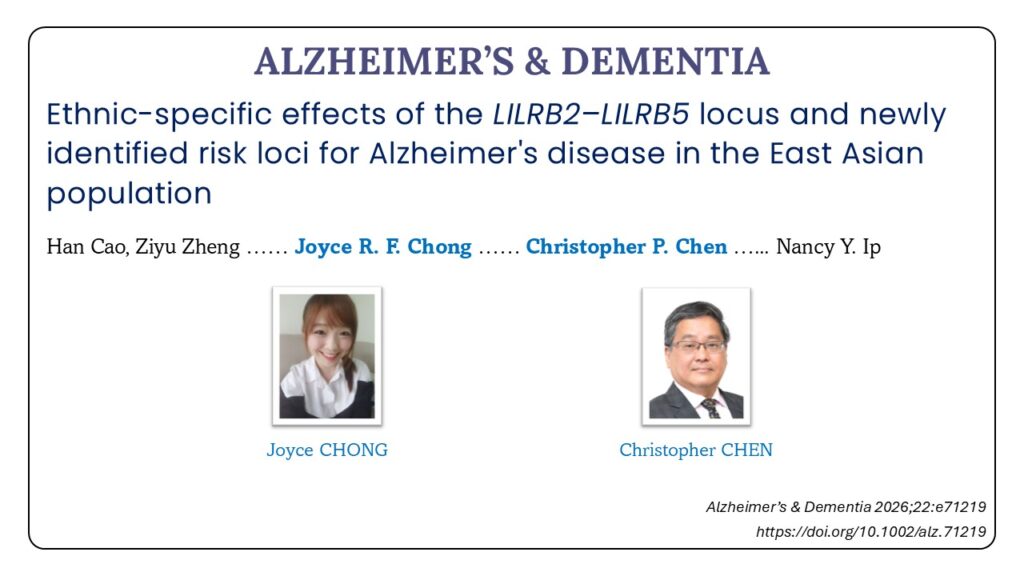
Genome-wide association studies have identified numerous Alzheimer’s disease (AD) susceptibility loci in European populations. However, the genetic architecture of AD in non-European populations remains underinvestigated.
We performed a genetic association study in East Asians (NÂ =Â 8514) to validate known AD loci and identify new susceptibility loci.
We identified LILRB2–LILRB5 as an AD susceptibility locus with ethnic-specific effects between Europeans and East Asians. The lead variant, rs587709-T, was associated with decreased AD risk and increased LILRB5 expression in Europeans. Conversely, in East Asians, the same allele was associated with increased AD risk and increased LILRB2 expression. Furthermore, genome-wide analysis identified TTC3 and FAM135A as candidate susceptibility loci for AD or cognition.
The results establish LILRB2–LILRB5 as a cross-ancestry AD-associated locus with ethnic-specific genetic mechanisms and reveal new susceptibility loci, extending the understanding of the genetic etiology of AD.
Full Article:Â https://alz-journals.onlinelibrary.wiley.com/doi/10.1002/alz.71219

c-Myc: Central regulator of autophagy and senescence in cancer Read More »


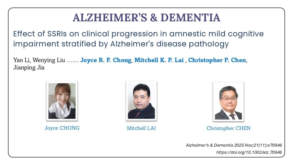
Introduction: This study examined whether selective serotonin reuptake inhibitors (SSRIs) treatment influenced cognitive trajectory and progression to Alzheimer’s disease (AD) dementia in amnestic mild cognitive impairment (MCI) patients, stratified by AD pathology.
Methods: Four hundred fifty-seven amnestic MCI participants in the ADNI database were analyzed. AD pathology was determined by baseline amyloid beta (Aβ) and tau positron emission tomography. Kaplan-Meier survival analysis and Cox proportional hazards models evaluated MCI-to-AD progression. Linear mixed models analyzed longitudinal cognitive trajectories, amyloid accumulation, and cortical thickness.
Results: SSRI treatment showed no significant effect on AD dementia progression (hazard ratio = 1.64, 95% confidence interval: 0.61 to 4.38) or cognitive trajectories, regardless of AD pathology. No significant differences in Aβ accumulation or cortical thickness were observed between SSRI users and non-users. External validation confirmed no significant SSRI effect on AD progression or cognitive decline.
Discussion: SSRI treatment was not associated with long-term cognitive effects in amnestic MCI, irrespective of underlying AD pathology.
Highlights: SSRI treatment was not associated with long-term AD dementia risk in MCI. SSRI treatment had no impact on long-term cognitive performance changes in MCI. SSRI treatment did not affect Aβ accumulation or cortical thickness in MCI. SSRIs had no effect on MCI progression, regardless of underlying AD pathology.
Full Article:Â https://pubmed.ncbi.nlm.nih.gov/41292493/

Global warming is expanding mosquito habitats and increasing mosquito-borne diseases. In tropical and sub-tropical regions, chikungunya virus (CHIKV) transmitted by Aedes mosquitoes has become a major concern due to the debilitating chronic joint disease it causes. Mosquito saliva contains bioactive factors that enhance viral infection, with sialokinin identified as a key contributor to vascular leakage and viral spread in mice. Here, we demonstrate that sialokinin binds to neurokinin receptors and restricts the activation of human myeloid cells. Mechanistically, sialokinin facilitates early viral dissemination, as evidenced by increased viral load in the contralateral footpad at 1 day post-infection, and significantly reduces circulating CD169+ monocytes while suppressing IFN-γ-producing T-cell-driven inflammation, as reflected by reduced joint footpad swelling in female CHIKV-infected mice. Clinically, patients with severe CHIKV disease exhibited higher levels of IgG antibodies against sialokinin, which correlated with higher viral loads and systemic inflammatory markers. Our findings highlight the multifaceted role of sialokinin in facilitating early viral dissemination and modulating host immunity during CHIKV infection. Given the growing threat of mosquito-borne diseases in a warming, disease-burdened world, targeting mosquito salivary factors like sialokinin could offer a novel therapeutic strategy to mitigate viral-induced inflammation and improve clinical outcomes.
Full Article:Â https://www.nature.com/articles/s41467-025-64468-x

Cortical cerebral microinfarcts are associated with brain atrophy in cross-sectional studies, with further investigation using longitudinal datasets being warranted. Moreover, little is known about their combined impact on cognition. This study aimed to establish the association between cortical cerebral microinfarcts and brain volume loss over time and explore whether they synergistically contribute to cognitive decline.
A total of 475 patients, aged 72.7 ± 7.9 years, were enrolled from a memory clinic cohort, who underwent neuroimaging and neuropsychological assessments at least twice over 5 years. Cortical cerebral microinfarcts and other cerebrovascular disease were assessed using 3-T MRI. Brain volumes were calculated semi-automatically using FreeSurfer. Cognitive function was assessed using a neuropsychological test battery including six domains. Linear mixed-effect models were utilized to examine the association between cortical cerebral microinfarcts and brain volume loss and their interaction on cognitive decline. Estimated marginal means were derived to plot global cognitive trajectories.
Cortical cerebral microinfarcts were associated with a greater decrease over 2 years in total brain volume [β = −1.94 (−3.07, −0.82) at Year 2, P-interaction with time < 0.001], grey matter volume [β = −1.00 (−1.69, −0.30) at Year 2, P-interaction = 0.002] and white matter volume [β = −0.95 (−1.54, −0.35) at Year 2, P-interaction < 0.001]. Brain volume loss was more pronounced in patients with multiple microinfarcts. Patients with high brain volume loss and cortical cerebral microinfarcts, particularly multiple microinfarcts, exhibited significantly lower global cognitive scores [single microinfarct: β = −1.83 (−2.68, −0.97) at Year 5, P-interaction with time < 0.001; multiple microinfarcts: β = −3.13 (−4.21, −2.05) at Year 5, P-interaction < 0.001]. The synergistic effects were more significant in the domains of executive function, memory, language and visuospatial function. Global cognitive trajectories revealed greater cognitive decline in patients with high brain volume loss and single or multiple microinfarcts, with the latter showing the steepest slope.
This study established a longitudinal association between cortical cerebral microinfarcts and brain atrophy progression, with higher microinfarct burden associated with more pronounced brain volume loss. Furthermore, cortical cerebral microinfarcts and brain atrophy showed synergistic effects on cognitive decline. These findings highlight the importance of investigating the role of mixed pathologies in the development of cognitive impairment and dementia in future research.
Full Article:Â https://academic.oup.com/brain/article/148/11/3924/8232732