Oncology
Early detection and staging of cancer is a key factor in a cancer patient’s long-term survival. Ongoing developments in whole body diffusion-weighted imaging and dynamic contrast-enhanced imaging will dovetail with development of PET-tracers for imaging specific tumour cell types. These will potentially improve early detection and diagnosis, and allow us to better monitor tumour progress and response to novel drug therapies.
Overall effectiveness of a therapy is crucially effected by the ability to institute prompt response-guided treatment changes. This requires development of imaging techniques that visualize anatomical and functional changes in the cancer cells early post-treatment.
At CIRC, we aim to develop imaging and analysis techniques that are targeted towards these goals of early detection and staging of cancer and early response assessment of therapy.
On-going Projects
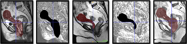
| Primary prostate cancer imaging with mpMRI, PET/CT and PET/MRI with focus on localization and grading |
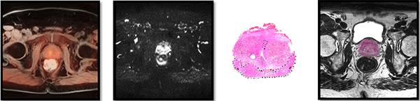 |
| The aims of this study are to find imaging modalities that are able to reliably localize cancer lesions within the prostate and to evaluate imaging parameters that correlate with ground truth tumor aggressiveness. Tracer kinetics, among others, are studied to separate aggressive from non-aggressive tumors. Biopsy results and histology data from specimen prostatectomy form the ground truth. This requires the challenging task of registering histology slices to the MR scan of the prostate to map the histological ground truth back to the MR images. |
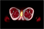
| Use of Hybrid Positron Emission Tomography/Magnetic Resonance in the treatment of Cervical Cancer |
 |
|
This project focusses on visualization of early tumour changes in cervical cancer patients who are undergoing concurrent chemotherapy, external beam radiotherapy and high dose rate brachytherapy. The goal is to detect early tumor response to brachytherapy treatment so that treatment parameters can be hand tuned based on ground truth tumor response instead of patient tolerance to radiation. The goal of this research is to develop a multi parametric imaging protocol and an image analysis framework to promptly analyse tumor after radiation administration. The image analysis is challenging due to the complex anatomical deformations introduced by the brachytherapy applicator that is inserted into the uterus during the treatment. |
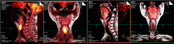
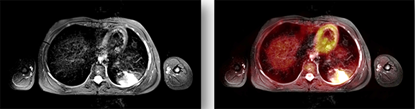
| Radiation response assessment by hybrid positron emission tomography/ magnetic resonance imaging for head and neck cancers |
|
|
|
Locoregional failure after radical radiation therapy can occur in a significant minority of patients. Typically, treatment response is assessed months after therapy due to confounding acute radiation therapy effects. This project investigates if hybrid positron emission tomography/ magnetic resonance imaging is able to visualize early tumour changes even during radiation therapy. The focus is on correlation of FDG-PET and MRI parameters within tumor sub-volumes to better understand difference(s) in head and neck tumor physiology such as vascular blood flow, permeability, apparent diffusion coefficient, among many others, between complete versus partial responders. |
| Is hybrid positron emission tomography magnetic resonance imaging (PET-MRI) an accurate modality for evaluation of rectal cancer patients after chemo-radiotherapy? |
|
|
|
The overall goal of this project is to explore the role of hybrid PET-MRI in evaluation of rectal cancer patients who have undergone chemo-radiotherapy. The aims are to evaluate accuracy of PET-MRI in re-staging and prediction of circumferential resection margin positivity post-therapy. This, in turn, will assess the correlation between PET-MRI functional parameters and treatment response. |
| The role of [18F]FDG PET/MRI in the diagnosis and management of febrile neutropenia- a pilot study |
 |
|
Febrile neutropenia is a common problem in children receiving chemotherapy for haematological-oncological conditions or undergoing haematopoietic stem cell transplantation. Neutropenia remains a diagnostic challenge because symptoms and signs can be absent or non-specific. The aim of this study is to assess whether whole-body imaging by PET/MRI with [18F]FDG is capable of detecting foci of infection and their specific features better than conventional imaging. |