Infectious Diseases
The use of positron emission tomography (PET) allows for visualisation of the functional processes occurring due to an infectious disease in the body. PET also provides the capability to assess the involvement of different sites of disease in the body. This has the potential to provide a more complete picture of organ involvement with disease than may be obtained from patient-reported symptoms, physical examination or from standard modalities of investigation.
Magnetic resonance imaging (MRI) provides highly detailed structural information in soft tissues and allows for visualisation of structures associated with a given disease. Fusing these structural images with the functional PET images gives a more complete picture of the behaviour of a disease process in the body and allows for an evaluation of treatment response to drugs as well as gaining a more complete picture of how a disease might affect various organs in the body.

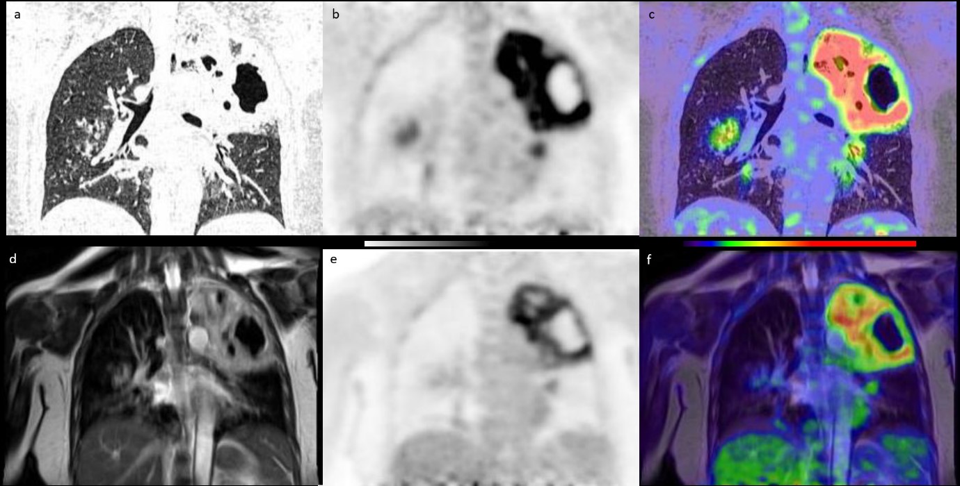
Figure 1 An example of TB visualisation in a patient using PET/CT and PET/MR data acquired at CIRC: top row shows a) CT, b) PET and c) fused PET/CT images; bottom row shows d) T2-weighted MRI, e) PET and f) fused PET/MR images. Images reproduced with permission from Thomas et al1.
Ongoing Projects
Programme of studies using 18F-FDG PET/MRI to image pulmonary tuberculosis and the response to drug treatment
There is a critical need for new biomarkers which can be used to evaluate the early response to tuberculosis (TB) drugs. Preliminary studies have shown that PET/CT appears to be useful in imaging pulmonary TB. PET/MRI has potential advantages over PET/CT for imaging TB in that it eliminates the radiation dose of CT (important when performing serial imaging to monitor treatment response) and can correct for motion in the PET scan. MRI itself shows good resolution of TB lesions and offers additional imaging possibilities. The combined modality may prove a major advance in TB imaging, and this is being explored as part of a programme of observational studies and clinical trials of TB in Singapore – Singapore Programme of Research Investigating New Approaches to Treatment of Tuberculosis, SPRINT TB.
Evaluation of 11C-acetate PET imaging as a novel approach to detecting pathology in pulmonary TB
Imaging using 11C-acetate PET in TB patients may be able to detect cells which are tolerant to antibiotics. This work may lead to greater understanding and identification of these persister cells within the pulmonary lesions of TB and ultimately assist in the development of biomarkers that may identify individuals with greater potential of relapse.
Evaluation of PET/MRI using a somatostatin analog tracer as a novel approach to detecting pathology in high risk TB-exposed contacts
PET/MRI imaging with somatostatin analog tracers has the potential to provide an imaging technique that allows identification of individuals with latent TB who may be at risk of progression to active TB. It is thought that 68Ga-DOTANOC imaging in latent TB will identify a subgroup of individuals with greater disease activity. This study will determine whether 68Ga-DOTANOC is more sensitive than 18F-FDG in identifying such disease in the lungs.
This work may lead to greater understanding of the spectrum of latent TB and ultimately assist in the development of biomarkers that may identify individuals with potential for progression to active disease.
Whole-body PET/MRI to evaluate the inflammatory response to Zika virus and dengue infection
There is a need to better understand the clinical spectrum of Zika and to relate this to the pathology – to understand the degree of organ involvement with Zika and in particular to understand the potential for neurological complications.
The scans that are done as part of this study are likely to provide detailed images of any inflammation or infection caused by the dengue or Zika virus. This study may add to the medical knowledge about Zika and dengue infection that may help to develop new ways of assessing and treating these infections that may benefit patients in the future.
1 B A Thomas et al. A comparison of 18F-FDG PET/MR with PET/CT in pulmonary tuberculosis. Nucl Med Comms.2017;38(11):971-978
