“Imaging for Health Symposium”
| DATE : | Wednesday, 11 November 2015 |
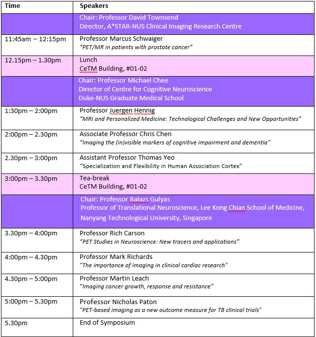
| TIME : | 11:45am – 5:30pm |
| VENUE : | Mary & Peter Fu Lecture Theatre (LT35), Centre for Translational Medicine, NUS |
Programme
Speakers
Professor Markus Schwaiger
Prof. Schwaiger has been Director of Nuclear Medicine at TUM’s rechts der Isar clinic since 1993. His main area of research is the use of multimodal imaging to visualize and quantify biological processes. His research involves the use of positron emission tomography (PET) in cardiology and oncology. He is Director of the German Research Foundation’s Collaborative Research Center 824, which deals with the use of molecular imaging for selecting and monitoring oncological therapies. The diagnosis and treatment of thyroid endocrine and neuroendocrine diseases is an area of special interest in the nuclear medicine department.
Prof. Schwaiger studied medicine at Freie Universität Berlin and Freiburg University. His career as a doctor and researcher has taken him to the UCLA School of Medicine (Los Angeles, USA) and the University of Michigan (Ann Arbor, USA), among other places, where he was professor of internal medicine until 1993. Prof. Schwaiger is a member of the Bavarian Academy of Sciences and the Leopoldina. He has an honorary doctorate from the University of Varna, Bulgaria
Professor Jurgen Hennig
Jürgen Klaus Hennig is a German chemistand medical physicist. Internationally he is considered to be one of the pioneers of Magnetic Resonance Imagingfor clinical diagnostics. He is the Scientific Director of the Department of Diagnostic Radiology and Chairman of the Magnetic Resonance Development and Application Center (MRDAC) at the University Medical Centre, Freiburg. In the year 2003 he was awarded the Max Planck Research Awardin the category of Biosciences and Medicine
Professor Rich Carson
Richard E. Carson, Ph.D. is Professor of Biomedical Engineering and Diagnostic Radiology at Yale University. He is Director of the Yale PET Center, and is also Director of Graduate Studies in Biomedical Engineering at Yale. He received his Ph.D. from UCLA in 1983 in Biomathematics and from then until 2005, Dr. Carson was a Senior Scientist in the PET program at the National Institutes of Health. Dr. Carson’s research interests include: 1) New algorithms for image reconstruction with PET, 2) Development of mathematical models for novel radiopharmaceuticals to produce images of physiological parameters, 3) Use of receptor-binding ligands to measure drug occupancy and dynamic changes in neurotransmitters by analysis of PET tracer signals, and 4) applications of PET tracers in clinical populations and preclinical models of disease, including neuropsychiatric disorders, diabetes, and cancer. Dr. Carson has published over 225 peer-reviewed papers, given over 100 invited lectures and is a member of the editorial board of the Journal of Nuclear Medicine, and the Journal of Cerebral Blood Flow and Metabolism. Honors include the Kuhl-Lassen award from the Brain Imaging Council of the Society of Nuclear Medicine, membership in the College of Fellows of the American Institute for Medical and Biological Engineering, the Sheffield Distinguished Teaching Award from the Yale School of Engineering, and the Edward J. Hoffman Memorial Award from the Computer and Instrumentation Council of the Society of Nuclear Medicine.
Professor Martin Leach
I have been motivated by translating developments in physics and related science to improve our understanding of disease and its treatment. My primary interest has been in developing and advancing magnetic resonance measurements to detect and diagnose cancer and to plan and evaluate cancer treatments.
With new treatments targeting cellular pathways that are central to cancer development, it has become increasingly important to develop imaging markers of target inhibition and to identify and characterise these in pre-clinical studies before moving these in to clinical studies. This involves identification of new biomarkers of pathways, and developing measurement and analysis methods to evaluate these biomarkers.
Following degrees in Physics and Applied Radiation Physics, I performed a research project on measuring body calcium by in vivo fast neutron activation analysis supervised by Professor John Fremlin in the Department of Physics at Birmingham. This involved working with the Nuffield Cyclotron, production of isotopes, development of analysis equipment and the study of inert gas behaviour in vivo.
I then joined the Joint Department of Physics at the Institute of Cancer Research to work on developing Computed Tomography (CT) scanners for radiotherapy planning, moving on to work with isotope imaging and a prototype Positron Emission Tomography (PET) system before developing the Magnetic Resonance (MR) programme with Janet Husband. The programme was initially clinically focussed and I developed applications of quantitative and functional imaging and MR spectroscopy to characterise cancer and its response to treatments. This led to many applications, including the MARIBS trial of MR for detecting breast cancer in women at high risk, which led to revised recommendations for management of high risk women by NICE and the American Cancer Society, resulting in the introduction of such screening in the cancer reform strategy.
During this period I also developed facilities for pre-clinical investigations, building a programme of metabolic investigation of new therapeutics and supporting development of a pre-clinical imaging programme. These facilities have resulted in measurements and analysis methods for functional MR have being developed and embedded in a range of clinical trials, including early stage trails of novel therapeutics.