Cardiology
Your heart is a muscle that pumps blood to your body, controlled by an electrical system. When the walls contract, blood is pumped into your circulatory system. Your heart is vital to your health. Without the heart’s pumping action, blood can’t move throughout your body. A healthy heart supplies your body with the right amount of blood at the rate needed to work well. If disease or injury weakens your heart, your body’s organs won’t receive enough blood to work normally.
Cardiovascular disease (CVD) is the major cause of death in western societies and represents 30 % of deaths in Singapore. It can be due to various causes from a non-healthy diet to a congenital disease. Diagnosing a CVD and planning the appropriate treatment if highly challenging. Usual diagnosis involve blood sampling and Echo imaging which gives important indicators, but fails in various diseases.
At CIRC we use state of the art MR, CT and combined PET-MR, PET-CT imaging equipment dedicated to research to answer fundamental questions so that today’s research becomes tomorrow’s clinical routine. Our team of cardiac MR specialists and image analysts allow to extend the studies possibilities for a wide range of applications.
Cardiovascular Magnetic Resonance (CMR)
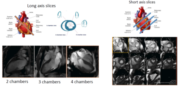
CMR offers many advantages over Echo with a higher image resolution, a 3D assessment of both ventricles, and its flexibility in the acquisition views so that no part of your heart stays invisible.
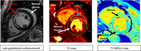
In addition, CMR offers structural information with or without the need for a contrast agent so that scar, fibrosis and edema be identified and accurately quantified.
On-going Projects
Using PET tracer [18F]NaF to differentiate between active and inactive calcium plaques in the coronaries arteries.
The goal of this study is to reproduce previously published results that use [18F]NaF PET-CT imaging to assess the activity of coronary plaques in order to predict the rupture of such plaques.
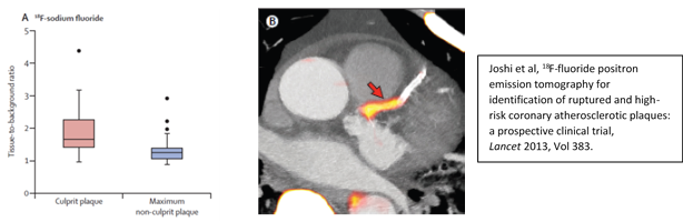
Using our PET-CT and PET-MR, we intend to assess the feasibility of [18F]NaF PET as a diagnostic tool to prevent plaque rupture as already reported, and combine PET activity with myocardial functional and structural information provided by MR imaging.
Combining MRS and CMR to guide the optimal timing of valve surgery for degenerative mitral valve regurgitation.
Degenerative Mitral Valve Regurgitation (DMR) is the most common underlying cause of severe mitral valve disease which may necessitate surgical valve repair or replacement. Defining the optimal timing for surgery is challenging in spite of advances in diagnostic imaging. CMR imaging is not the primary diagnostic method for DMR but may offer information on function, structure and flow potentially improving grading of the severity of disease and help guide the timing of intervention. In addition, 31P MRS can provide energetic and metabolic information that standard echo cannot provide, for the assessment of severity of DMR-related changes in ventricular structure.
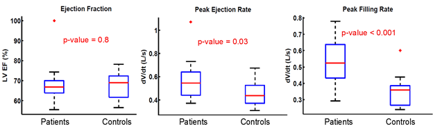
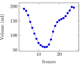
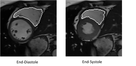
At CIRC, assessment of left ventricular function is performed semi-automatically on the entire cardiac cycle. This allows to consider standard parameters as well as novel ones that are not routinely measured by radiologists, for instance rate of ejection and filling, which could be significant indicators of a particular disease.
Studying the impact of pharmacological therapy on plaque vulnerability and temporal changes on STEMI patients.
In acute ST-elevation MI (STEMI), myocardial injury induces an intense inflammatory and fibrotic response that extends well beyond the infarct border zone. This leads to dilatation and distortion of the left ventricle, also known as adverse left ventricular (LV) remodeling. We plan to study the correlation between prolonged thrombin activity and adverse ventricular remodeling following STEMI. 100 patients are under recruitment for this study.
Comparing telemedicine patient care to standard usual care on high risk post-Myocardial Infarction patients.
An unmet need exists for improved post-MI strategies to avoid left ventricular remodeling and heart failure. The current study aims to compare patients who were monitored under telemedicine versus standard care group to determine the outcomes by comparing the difference in LVEDV, LVESV, LVEF with CMR scan as surrogate endpoint both at baseline and end of 6 month’s telemedicine monitoring.
More than 300 patients are under recruitment for this study.
Image Analysis
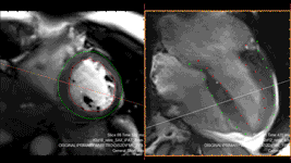
At CIRC, we perform automatic analysis of every cardiac sequences, including 4D left and right ventricle segmentation, automatic scar, edema and fibrosis delineation, as well as automatic flow analysis and multi-modality registration.
Left ventricular segmentation (using Segment/Medviso)
Right ventricular segmentation (in-house software)
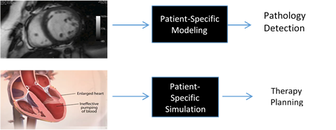
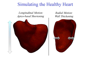
Patient-specific simulation
In order to predict cardiac therapy and therefore diminish the number of heart failures, many research projects are trying to understand the heart behavior, to model and simulate a cardiac cycle in healthy cases as well as pathological ones. The goals are to extract intrinsic mechanical parameters to help pathology detection, and to try several therapies using computerized models in order to select the best possible therapy for a given patient.