
Issue 41 / February 2022
Insights
Slaying a Serial Killer –
The Decade-long Quest by an NUS and Glaxosmithkline Team
A decade-long unpredictable journey of malaria cell death research—from bench to industry and back.
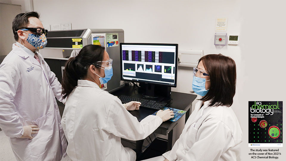
n October 2021, a team at the NUS Laboratory of Molecular and Cellular Parasitology realised a cherished dream of publishing a major study based on a collaboration with GlaxoSmithKline’s (GSK) Global Health Discovery Incubator Unit1. The project involved the use of an advanced imaging protocol developed by the group to screen GSK’s drug library for drugs that kill malaria parasites through disruption of the cell’s calcium balance. The NUS team performed the screen, while the GSK team in Spain carried out detailed characterisation of the effective compounds. The method resulted in the identification of potent antimalarials with a unique mode of killing action, providing the global malaria community with invaluable starting points for drug development. The featuring of the study on the cover of ACS Chemical Biology was certainly a bonus.
The origins of the work go back a decade, when an eager PhD student published, in 2010, a seminal study on drug-mediated suicide in malaria parasites.
Cellular suicide, also called apoptosis or programmed cell death (PCD), is a genetically encoded feature of multicellular organisms and serves the vital function of deleting unwanted, damaged or infected cells, so as to preserve the health and integrity of the organism. In contrast, the field of PCD in single-cell organisms was, and continues to be, controversial.
The existential question is ‘why would a single cell kill itself, as this would result in the death of the entire organism’. Yet, there are numerous reports of single cell organisms that exhibit molecular and cellular features of PCD, so much so that there exists an international journal dedicated to the study of PCD in unicellular organisms (https://microbialcell.com/). The malaria parasite, a single-cell eukaryote, was reported to exhibit some features of cellular suicide when exposed to specific antimalarials. However, other reports concluded that no such features were discernible.
The malaria parasite, a single-cell eukaryote, was reported to exhibit some features of cellular suicide when exposed to specific antimalarials.
Dr Ch’ng Jun Hong, then a PhD student with our department, reconciled the controversy by showing that the suicidal signatures were evident depending on the concentration of the drug used2. In quick succession, he reported that these death signatures occurred early during drug exposure and involved calcium ion leakage from a specific cell compartment3. This made sense. Cellular calcium, as an ion, is vital for cellular processes, but is highly reactive. Hence, cells need to store calcium in specific compartments (lysosomes, mitochondria, etc.) to prevent activation of calcium responsive enzymes, leading to cell death. Some antimalarials cause calcium ions to spill out of their stores, activating enzymes that essentially digest the parasite from within. Importantly, he went on to report that these calcium-mediated cell death features were physiologically relevant and could be observed in mouse models of malaria infection4.
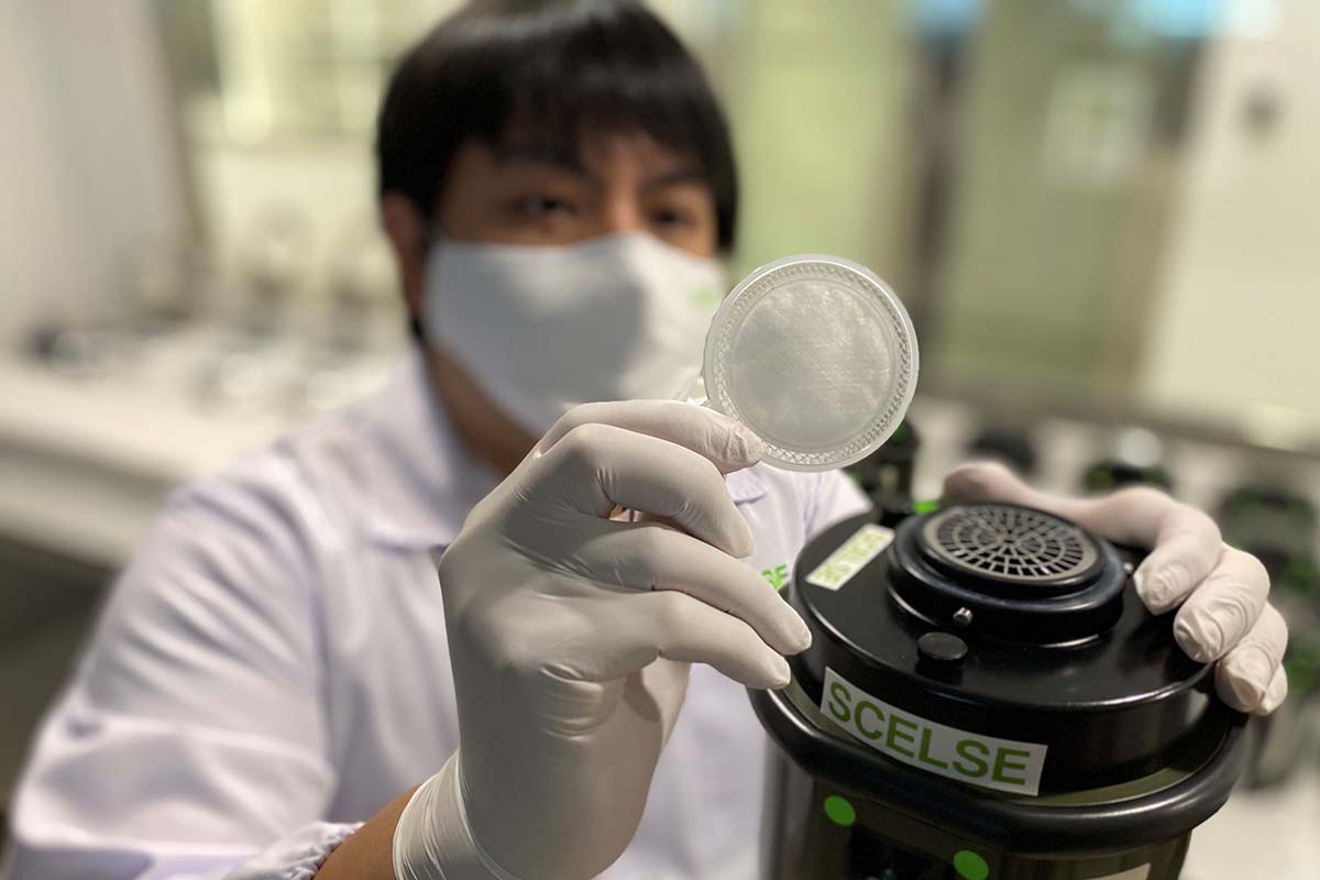
Around this time, a new type of imaging technology was gaining popularity. The flow-based imaging platform exploited improvements in computer processor speeds to enable split-second image capture and analysis of several thousand cells flowing past a high-speed camera. Because we could measure calcium location within a cell using calcium-sensitive fluorescent probes, this imaging technology seemed ideal for screening drugs that disrupted calcium balance in large numbers of parasites; malaria parasites that succumbed to such drugs would reveal images of calcium spilling out of their stores and into the cytosol, on a large scale. This was achieved on a small panel of drugs5 and served as a proof of concept for screening a larger collection of compounds.
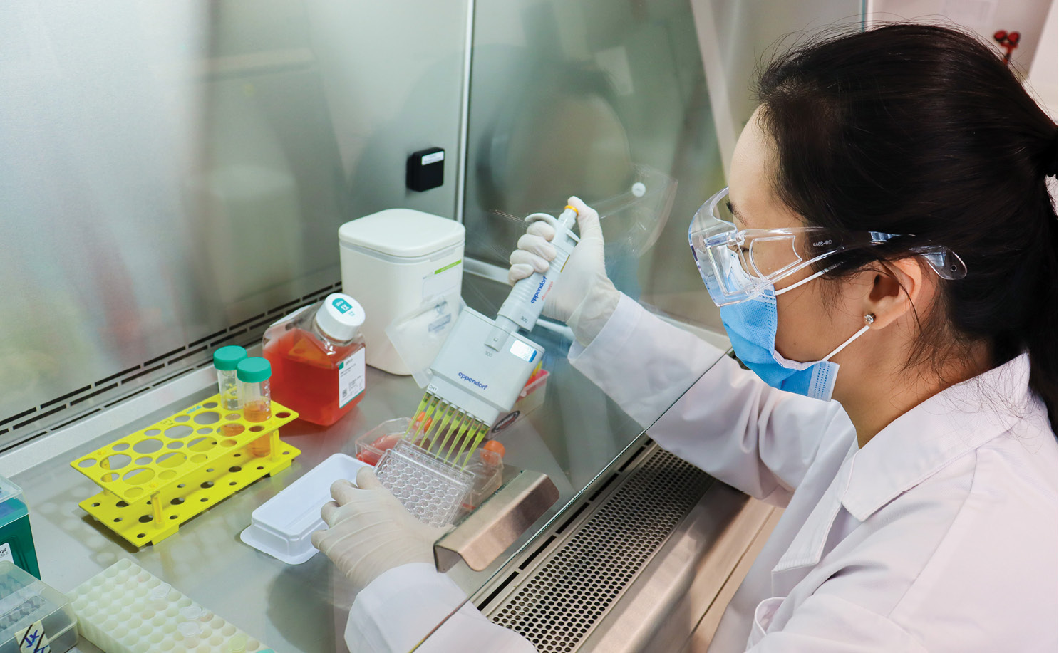
Dr Wanni Chia (first author, post-doctoral researcher) doing malaria cell culture.
In 2014, the Department of Microbiology and Immunology purchased the latest flow-based imaging platform (Amnis ISX MKII), which enabled many more compounds to be screened in a short span of time. Fortuitously, and around this period, I met with GSK Singapore’s strategy and partnership development manager, Dr Yasuji Matsuoka, during a networking session at an enterprise convention. He highlighted our capability to GSK’s malaria team, and we were subsequently engaged to screen their antimalarial library of compounds for calcium disrupting effects.
Our GSK-Spain collaborator was Dr Maria de Gracia Gomez‑Lorenzo, an affable, professional and responsive investigator. Over S$100,000 of research funds were allocated, which allowed us to hire Dr Chia Wanni as a postdoctoral fellow, to manage the project for a year. The methodology was further refined during her time on the project, which was published in a leading methods journal6. Around this time, Dr Tong Jiexin, then a PhD student, validated the utility of the method by screening a different compound library7. Her work branched out into several smaller projects, one of which led to an unexpected finding, which I hope to share on another occasion.
Another study is presently undertaken by our current PhD student, Claudia Carrera Bravo, who is seeking to shed light on the cellular and immunological consequences of calcium destabilisation in drug-treated malaria parasites, in collaboration with Prof Laurent Renia from A*STAR. So that’s where we are today. It has been a long but productive journey, filled with twists and turns.
Looking Back at My Decade-long Research Journey
Research usually takes on an unpredictable trajectory. Dead ends are common, but sometimes, the stars align, and things work out. The journey becomes clearer on hindsight, so it is good to keep an open mind, and leverage on opportunities when they present themselves.
Impactful work, especially in the biomedical domain, doesn’t happen overnight. It takes years of committed and strategic focus for projects to bear fruit.
You need a talented research workforce to deliver high quality data and publications. Drs Ch’ng Jun Hong, Chia Wanni and Tong Jiexin were instrumental to the success of our projects.
Networking is crucial for expanding our opportunities. My chance meeting with Dr Matsuoka gave birth to an industry collaboration that most academics would dream of. When a pharmaceutical giant takes notice of your work, it is proof that your research is impactful and relevant.
Never, never, give up. Our featured paper¹ was rejected by several journals, partly due to the controversial nature of PCD in malaria. In recent months, our new colleagues, Drs Rajesh Chandramohanadas and Trang Chu, both malaria experts, joined our efforts to reshape the project and add new data to further strengthen our work. This strategy worked, and the rest is history.
-
Chia W, Gomez-Lorenzo MG, Castellote I, Tong JX, Chandramohanadas R, Thu Chu TT, Shen W, Go ML, de Cozar C, Crespo B, Almela MJ, Neria-Serrano F, Franco V, Gamo FJ, Tan KSW (2021) High-content phenotypic screen of a focused TCAMS drug library identifies novel disruptors of the malaria parasite calcium dynamics. ACS Chem. Biol. 16:2348-2372.
-
Ch’ng JH, Kotturi SR, Chong AGL, Lear MJ and Tan KSW (2010) A programmed cell death pathway in the malaria parasite Plasmodium falciparum has general features of apoptosis but is mediated by clan CA proteases. Cell Death Dis. 1 e26.
-
Ch’ng JH, Liew K, Goh ASP, Sidhartha E, Tan KSW (2011) Drug-induced permeabilization of parasite’s digestive vacuole is a key trigger of programmed cell death in Plasmodium falciparum. Cell Death Dis. 2 e216.
-
Ch’ng JH, Lee YQ, Gun SY, Chia WN, Chang ZW, Wong LK, Batty KT, Russell B, Nosten F, Renia L and Tan KSW (2014) Validation of an alternative antimalarial mechanism of chloroquine for clinical use against malaria. Cell Death Dis. 5 e1305.
-
Lee YQ, Goh ASP, Ch’ng JH, Nosten FH, Preiser PR, Pervaiz S, Yadav SK and Tan KSW (2014) A high-content phenotypic screen reveals the disruptive potency of quinacrine and 3’,4’-dichlorobenzamil on the digestive vacuole of Plasmodium falciparum. Antimicrob. Agents Chemother. 58(1): 550-558.
-
Chia WN, Lee YQ, Tan KSW (2017) Imaging flow cytometry for the screening of compounds that disrupt the Plasmodium falciparum digestive vacuole. Methods 112:211-220.
-
Tong JX, Chandramohanadas R, Tan KSW (2018) High-content screening of MMV Pathogen Box for Plasmodium falciparum digestive vacuole disrupting molecules reveals valuable starting points for drug discovery. Antimicrob. Agents Chemother. 62(3): e02031-17.