Abstract
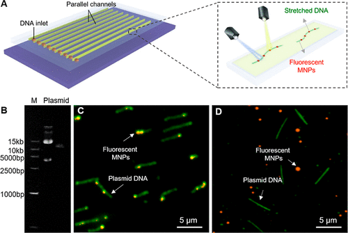
As the key player of a new restriction modification system, DNA phosphorothioate (PT) modification, which swaps oxygen for sulfur on the DNA backbone, protects the bacterial host from foreign DNA invasion. The identification of PT sites helps us understand its physiological defense mechanisms, but accurately quantifying this dynamic modification remains a challenge. Herein, we report a simple quantitative analysis method for optical mapping of PT sites in the single bacterial genome. DNA molecules are fully stretched and immobilized in a microfluidic chip by capillary flow and electrostatic interactions, improving the labeling efficiency by maximizing exposure of PT sites on DNA while avoiding DNA loss and damage. After screening 116 candidates, we identified a bifunctional chemical compound, iodoacetyl-polyethylene glycol-biotin, that can noninvasively and selectively biotinylate PT sites, enabling further labeling with streptavidin fluorescent nanoprobes. With this method, PT sites in PT+ DNA can be easily detected by fluorescence, while almost no detectable ones were found in PT– DNA, achieving real-time visualization of PT sites on a single DNA molecule. Collectively, this facile genome-wide PT site detection method directly characterizes the distribution and frequency of DNA modification, facilitating a better understanding of its modification mechanism that can be potentially extended to label DNAs in different species.
Read More: https://pubs.acs.org/doi/abs/10.1021/acs.analchem.2c01752


