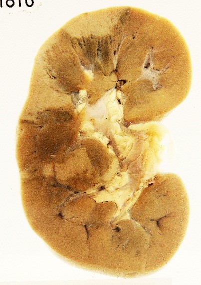
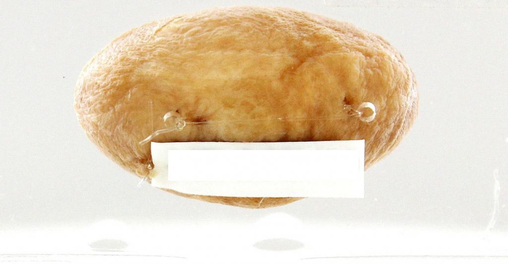
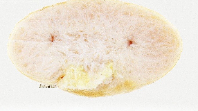
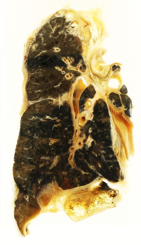
Exercise: Take a look at these pictures, and practise describing them systematically – organ, orientation, main pathology (diffuse or localised), describe specific features.
Click on the DESCRIPTION boxes to see how they may be described.
View the VIDEO descriptions as well .
Case 1: KIDNEY

Click HERE to view video.
If you can't play the video, watch it on Youtube: https://youtu.be/mIYihwgw2dQ
Case 2: Skin Lesion (External View and Cut Section)


Case 3: Lung

Click HERE to view video.
If you can't play the video, watch it on Youtube: https://youtu.be/YW9xe0mpzKs
Note: Please do take a minute or two to fill in your comments below, e.g. Is this useful? Would you like more? Though not required, feel free to include your contact details as well, if so inclined!
Created by: Dr Nga Min En, Department of Pathology, NUS Yong Loo Lin School of Medicine, Singapore
Acknowledgements:
Mr Muhamad Aidil Bin Johari, for technical advice
Mr Arif Uzair (Class of 2016) for his valued inputs
More talking pots/slides in: Virtual Pathology Museum and YouTube (Pathweb Teacher).
View our Catalogue of Virtual Pathology specimens and talking pots/slides.
7 Comments
Leave a Comment
You must be logged in to post a comment.

This is really useful, especially the video explanations! Thank you!
Hi I am glad you find it so! There are many topic overviews in Pathology Demystified too, which I hope you will find useful as well, to lessen your pain!
Happy learning.
Dr Nga
This is very nice and useful, very useful for self directed learning for students, the quality of both gross and microscopic images are excellent
Yes the videos are really good. I wish I had seen this sooner.
Thank you for the videos. It is very understandable 🙂
Thank you, it is useful and easier to practice!
This whole guide series is extremely helpful. Thank you very much.