Multimodal Brain Imaging Analysis
A vast proportion of the population will be affected by neuropsychiatric conditions during their lifetime, including stroke, dementia and psychiatric illnesses. However, the causes and underlying changes are often unclear.
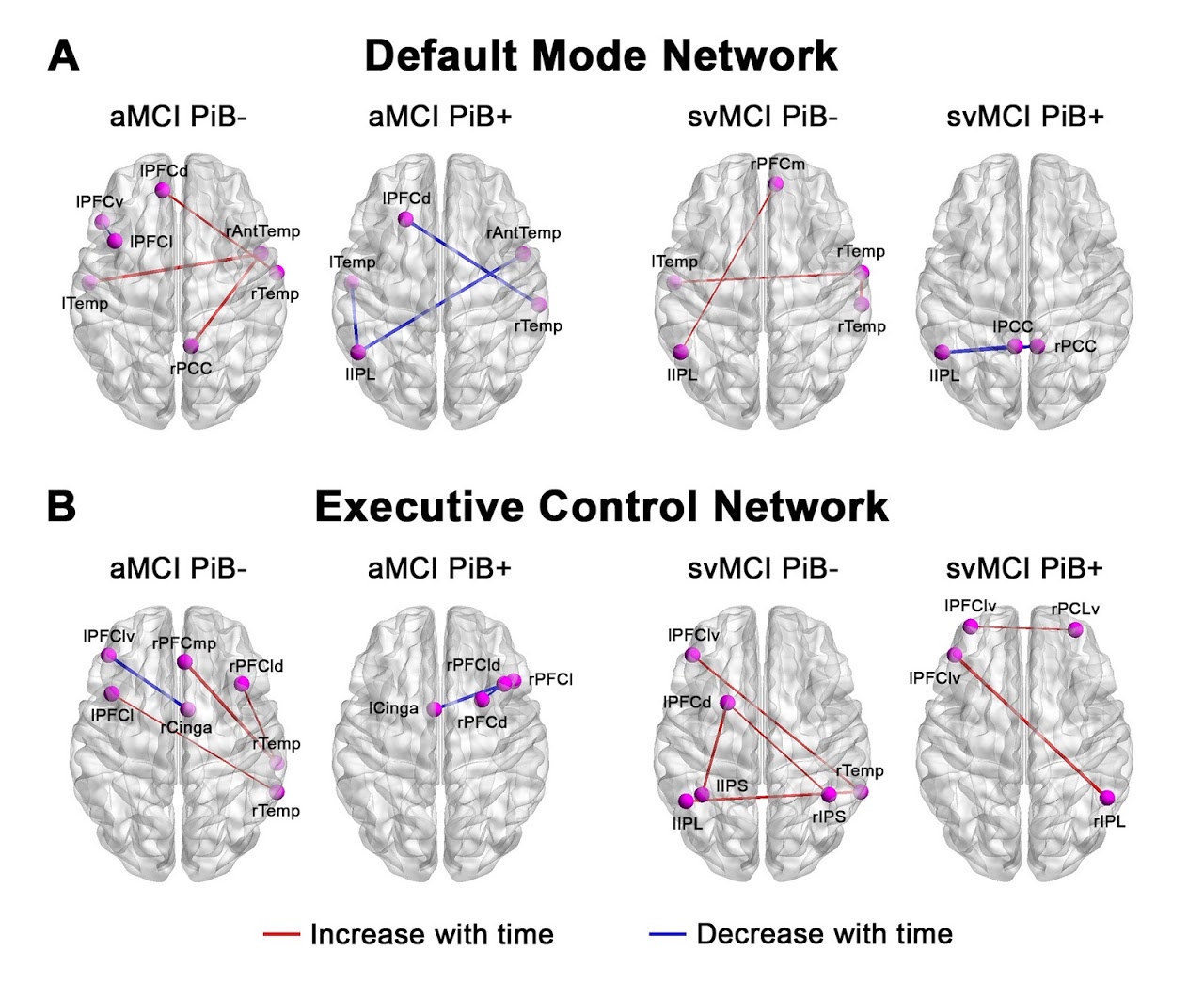
At TMR we have many studies using brain imaging (e.g., MRI, diffusion MRI, EEG) to understand and predict the development of neurological/psychiatric conditions, as well as to monitor potential treatments. We also support projects trying to understand healthy brain network organization, development, and aging.
The TMR leadership team is internationally recognized for their work on brain imaging and analysis: Dr. Zhou was the Secretary of the Organization for Human Brain Mapping (OHBM) Council, Dr. Chee is a Fellow of the OHBM and Dr. Yeo is a recipient of the OHBM Early Career Investigator Award 2019.
Some of our most commonly used imaging techniques are listed below.
| Measurement | Acquisition | Analysis |
| Spontaneous Brain Activity | Resting-state fMRI | Functional connectivity |
| Regional Brain Activity | Task-fMRI (MS-EPI) | Automated analysis of task-based fMRI data. |
| Brain Volumes | T1w TFE | Automated analysis of cortical parcellations enabling measurement of regional brain volumes and other morphological measures |
| Edema | FLAIR | Automated analysis of white matter hyperintensities, often seen in ischaemia and stroke. |
| Microbleeds | SWI | Delineation of regions of altered susceptibility due to microbleeds. |
| Perfusion | ASL (pCASL/GRASE) | Automated analysis for cortical perfusion. Partial volume correction possible. |
| Neuronal Orientation and dispersion | NODDI/ DTI | Semi-Automated analysis producing images of neuronal orientation and dispersion. |
| Water diffusion | Diffusion MRI | Tractography, axon and myelination, free water estimation |
| Vessel imaging | SPACE/TOF | Identification of vessel narrowing. |


