Digital transformation of histology – A new trend in medical teaching

Submitted: 13 September 2023
Accepted: 4 March 2024
Published online: 2 July, TAPS 2024, 9(3), 64-66
https://doi.org/10.29060/TAPS.2024-9-3/CS3138
Jayabharathi Krishnan, Sara Kashkouli Rahmanzadeh & S. Thameem Dheen
Department of Anatomy, Yong Loo Lin School of Medicine, National University of Singapore, Singapore
I. INTRODUCTION
In preclinical years, histology, which is the study of the microscopic structures of tissue and organs, aids students in understanding the normal morphology of cell and tissue organisation in organs and differentiating their pathological changes (Hussein & Raad, 2015). The study of histology is important as it provides the fundamental basis of anatomical knowledge. Students have adapted to a new learning environment, particularly after the COVID-19 outbreak, by utilising autonomous learning strategies, including online and digital learning, as histology requires visual interpretation that is developed by continuous practice (Yohannan et al., 2019). Given this, we have created a virtual histology platform using our existing tool: the National University of Singapore – Human Anatomy Learning resOurce (NUS-HALO). NUS- HALO is an online platform with digital images and videos and has emerged as a novel tool in transforming anatomy teaching and learning. By integrating cutting-edge, high-definition histology images and relevant learning materials, the histology component of NUS-HALO offers a platform that aids students to excel in histology (Darici et al., 2021).
The NUS-HALO platform aids student learning of histology. Histology resources are organised systematically, along with pertinent teaching resources and explanations, to help students better comprehend each histological slide. Furthermore, during their third year of medical school, when students are introduced to pathology, they must use their earlier understanding of normal histology to identify pathological changes.
II. METHODS
Our team included three technical and five academic staff members. The digitisation of histology sections, selected from our existing collection in the Department, was done using Aperio software, and the digital images were saved on a server to be accessed later for teaching. Overall, images from 160 histology slides comprising 13 organ systems were digitised, each taking about 90 minutes to digitise. These images were clustered with the previously saved images (200 images from seven organ systems) and selected for NUS-HALO’s histology arm. The histology slides were carefully chosen to obtain low- and high-magnification images. The images were labelled to give students a clear understanding of each organ system and its critical features.
III. DISCUSSION
NUS-HALO offers a platform that aids student learning of histology. Histology resources are organised systematically, along with pertinent teaching resources and explanations, to help students better comprehend each histological slide. Furthermore, during their third year of medical school, when students are introduced to pathology, they must use their earlier understanding of normal histology to identify pathological changes.
HALO’s histology resources can be seamlessly integrated with what students learn during their anatomy and physiology classes. The use of this tool allows for a holistic understanding where students are able to correlate the microscopic histological structures with macroscopic anatomical features and physiological functions. Informal feedback that has been obtained from both staff and students has been overwhelmingly positive, highlighting the ease of use and quality of the resources available on the platform. A notable outcome has been the informal feedback received by students, stating that the platform has aided their examination preparation. However, continued and more formal gathering of feedback is essential for the platform’s ongoing improvement.
Future enhancements of the platform include using more diverse slide samples, and more interactive elements such as self-evaluation guides to enhance student’s experience and the effectiveness of NUS-HALO. Self-evaluation guides that are currently being considered include identification exercises, where students name structures on slides, and interpretive questions that can test their understanding of how histological changes might relate to pathological conditions. These tools will reinforce learning and enable students to track their progress.
A. Pedagogical Framework of Digital Histology on NUS-HALO
The resources on the NUS-HALO webpage were organised as follows:
1) Categorisation of Histology Images:
Images were organised based on the organ system they belong to (e.g., respiratory, digestive). Each image was annotated with labels and identification markers highlighting fundamental structures and features.
2) Integration of Teaching Resources:
Short notes describing salient features of the sections were embedded alongside the corresponding histology images to provide students with further explanations.
3) Navigation and User Interface:
The resources were organised to facilitate easy navigation, with a search function, intuitive m menus, and clear headings.
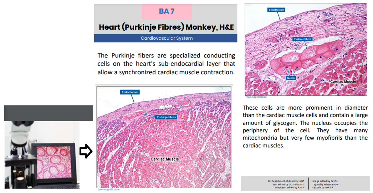
Figure 1: Showing high-quality images captured and uploaded for student access (leftmost). Image showing information available to students when they select digitised slides.
IV. CONCLUSION
The advent of computer-aided digital media and images has significantly impacted medical education, including image-intensive histology. Digitising histology slides appears cost-effective as it reduces the need for microscope maintenance and preparation of glass slides when damaged and manpower costs. This tool serves as an additional learning resource that students can access in conjuction with their existing histology lectures or practical lessons.
In the future, digital histology can be enhanced by incorporating augmented and virtual reality and artificial intelligence to provide students with an enhanced, immersive, and interactive learning experience.
Note on Contributors
Dr. Jayabharathi Krishnan, Dr. Sara Kashkouli Rahmanzadeh, and Professor S. Thameem Dheen are content experts on the Histology aspect of NUS-HALO. All authors contributed equally to this manuscript.
Acknowledgement
The authors would like to acknowledge the technical staff: Ms . Pan Feng, Ms. Bay SL, Ms. Rebecca Auw, and Mr. Low CP from the Department of Anatomy, Yong Loo Lin School of Medicine, for their technical support. The authors would sincerely like to thank the Department of Pathology for contributing to developing the digitalised images.
Funding
The authors did not receive any funding for this study.
Declaration of Interest
The authors do not have any conflict of interest.
References
Darici, D., Reissner, C., Brockhaus, J., & Missler, M. (2021). Implementation of a fully digital histology course in the anatomical teaching curriculum during covid-19 pandemic. Annals of Anatomy – Anatomischer Anzeiger, 236, Article 151718. https://doi.org/10.1016/j.aanat.2021.151718
Hussein, I. H., & Raad, M. (2015). Once upon a microscopic slide: The story of histology. Journal of Cytology & Histology, 6(6), Article 1000377. https://doi.org/10.4172/2157-7099.1000377
Yohannan, D. G., Oommen, A. M., Umesan, K. G., Raveendran, V. L., Sreedhar, L. S., Anish, T. S., Hortsch, M., & Krishnapillai, R. (2019). Overcoming barriers in a traditional medical education system by the stepwise, evidence-based introduction of a modern learning technology. Medical Science Educator, 29(3), 803–817. https://doi.org/10.1007/s40670-019-00759-5
*DR K JAYABHARATHI
Department of Anatomy
Yong Loo Lin School of Medicine
MD10, 4 Medical Drive
Singapore 117594
Email: antkj@nus.edu.sg
Announcements
- Best Reviewer Awards 2025
TAPS would like to express gratitude and thanks to an extraordinary group of reviewers who are awarded the Best Reviewer Awards for 2025.
Refer here for the list of recipients. - Most Accessed Article 2025
The Most Accessed Article of 2025 goes to Analyses of self-care agency and mindset: A pilot study on Malaysian undergraduate medical students.
Congratulations, Dr Reshma Mohamed Ansari and co-authors! - Best Article Award 2025
The Best Article Award of 2025 goes to From disparity to inclusivity: Narrative review of strategies in medical education to bridge gender inequality.
Congratulations, Dr Han Ting Jillian Yeo and co-authors! - Best Reviewer Awards 2024
TAPS would like to express gratitude and thanks to an extraordinary group of reviewers who are awarded the Best Reviewer Awards for 2024.
Refer here for the list of recipients. - Most Accessed Article 2024
The Most Accessed Article of 2024 goes to Persons with Disabilities (PWD) as patient educators: Effects on medical student attitudes.
Congratulations, Dr Vivien Lee and co-authors! - Best Article Award 2024
The Best Article Award of 2024 goes to Achieving Competency for Year 1 Doctors in Singapore: Comparing Night Float or Traditional Call.
Congratulations, Dr Tan Mae Yue and co-authors! - Best Reviewer Awards 2023
TAPS would like to express gratitude and thanks to an extraordinary group of reviewers who are awarded the Best Reviewer Awards for 2023.
Refer here for the list of recipients. - Most Accessed Article 2023
The Most Accessed Article of 2023 goes to Small, sustainable, steps to success as a scholar in Health Professions Education – Micro (macro and meta) matters.
Congratulations, A/Prof Goh Poh-Sun & Dr Elisabeth Schlegel! - Best Article Award 2023
The Best Article Award of 2023 goes to Increasing the value of Community-Based Education through Interprofessional Education.
Congratulations, Dr Tri Nur Kristina and co-authors! - Best Reviewer Awards 2022
TAPS would like to express gratitude and thanks to an extraordinary group of reviewers who are awarded the Best Reviewer Awards for 2022.
Refer here for the list of recipients. - Most Accessed Article 2022
The Most Accessed Article of 2022 goes to An urgent need to teach complexity science to health science students.
Congratulations, Dr Bhuvan KC and Dr Ravi Shankar. - Best Article Award 2022
The Best Article Award of 2022 goes to From clinician to educator: A scoping review of professional identity and the influence of impostor phenomenon.
Congratulations, Ms Freeman and co-authors.
