Question 1:

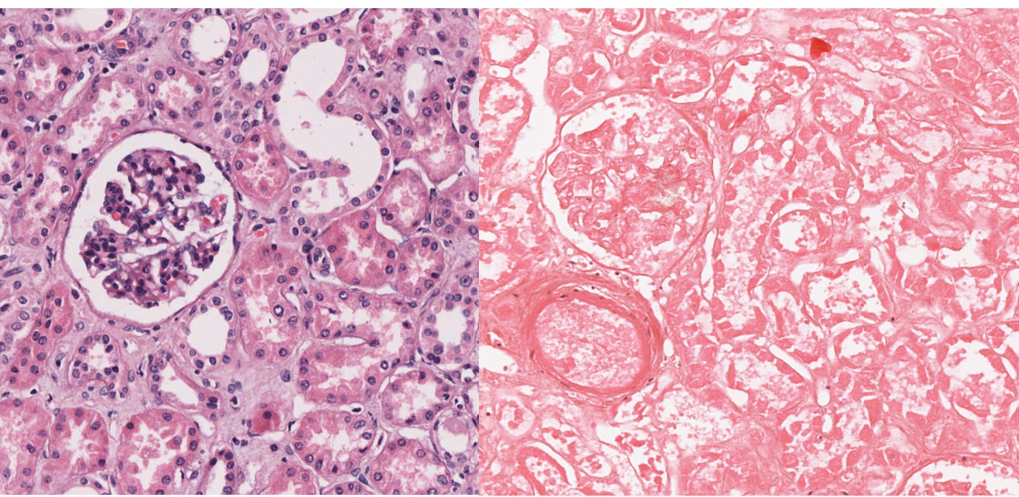
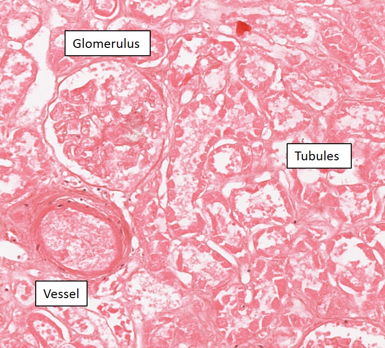
a. What organ do the microscopic pictures feature?
b.Describe the microscopic changes seen in the picture on the RIGHT.
c. What is the pathologic process?
d. What is the diagnosis and the likely cause?
Question 2:

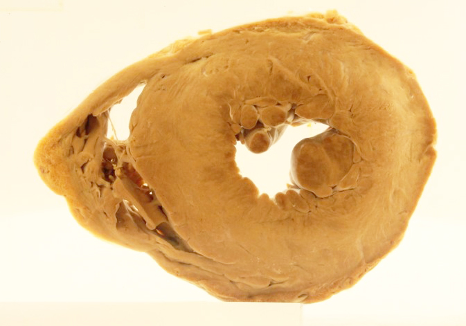
57 year old man. Referred to cardiologist after routine health screening.
a. Describe the main abnormality seen and give the diagnosis.
b. What specific type of cellular response does this represent? What is a likely cause in this patient?
c. What would you expect to see on microscopic examination of the abnormal area?
Question 3:

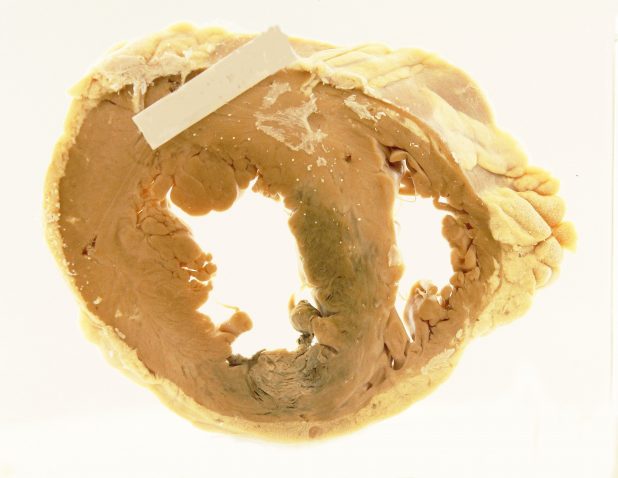
63 year old man. Complained of severe chest pain and sweating before collapsing.
a. Describe the main abnormality seen and give the diagnosis.
b. What specific type of cellular response does this represent?
c. Describe what you are likely to see under the microscope.


