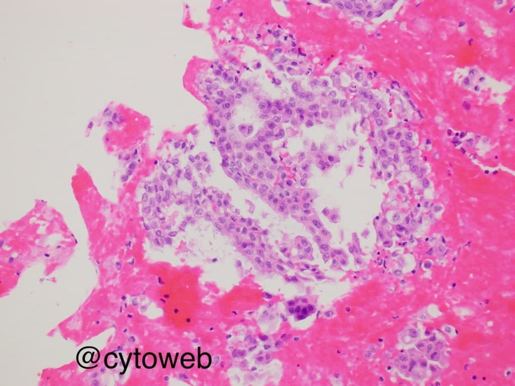
Secretory carcinoma
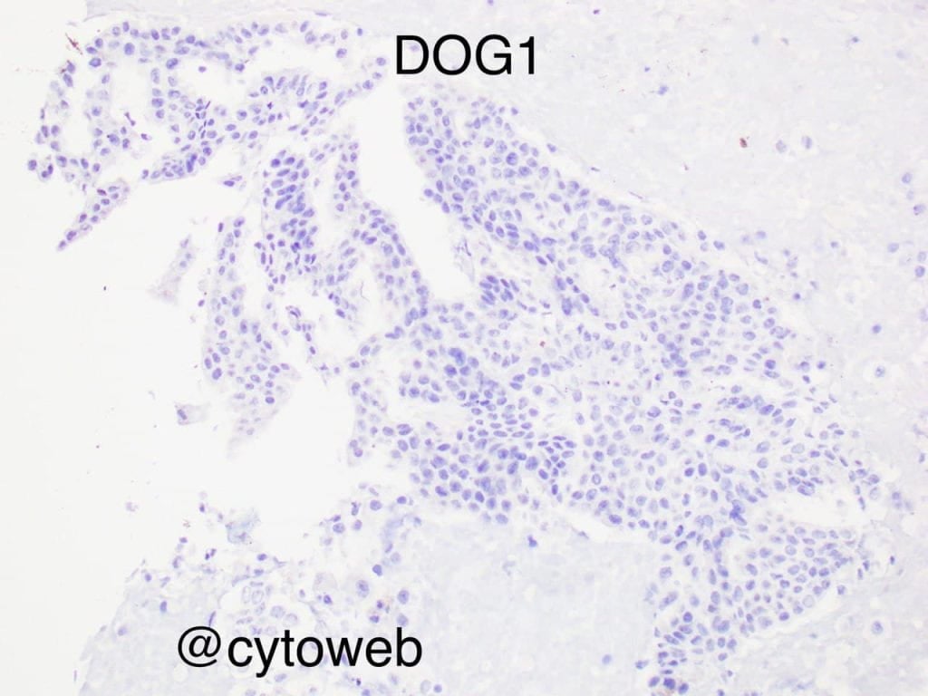
A definitive morphologic cytologic diagnosis of secretory carcinoma is difficult as there is often some degree of cytomorphologic overlap with acinic cell carcinoma, mucoepidermoid carcinoma (low grade) and intraductal carcinoma.
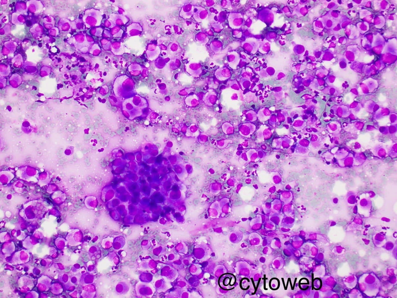
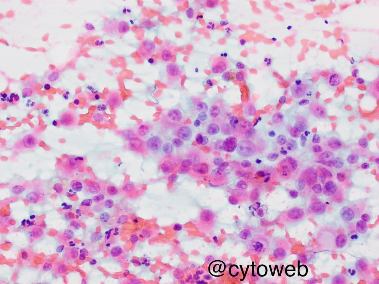
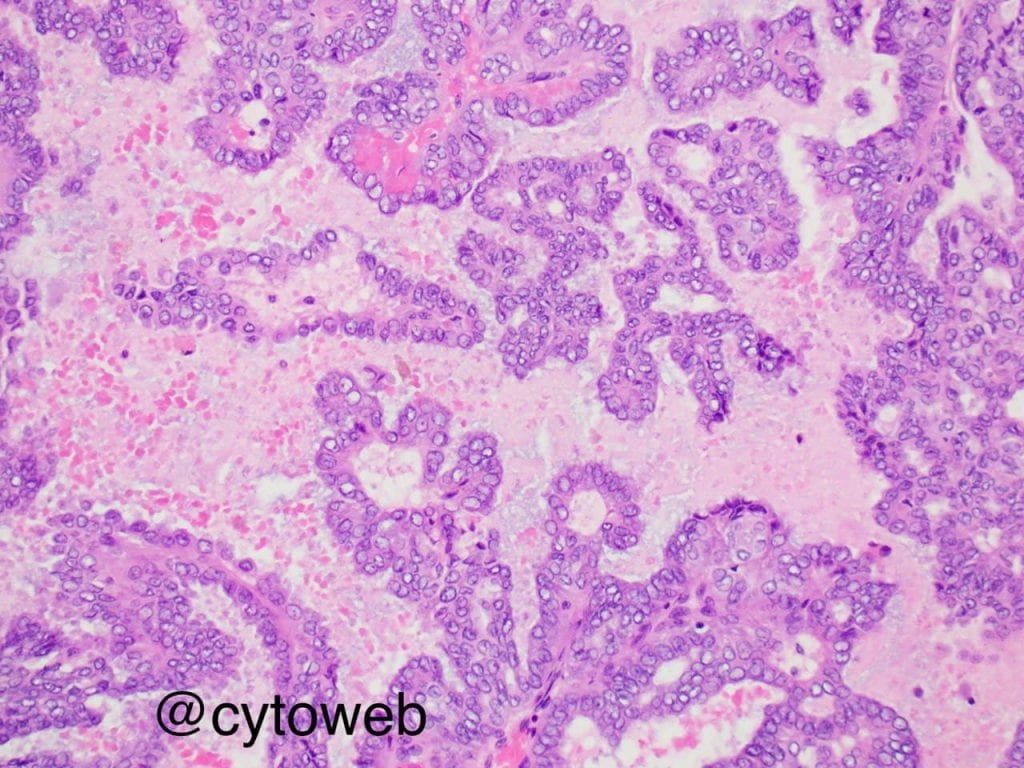
The salient, but non-specific, cytomorphologic features of secretory carcinoma include the presence of epithelioid cells arranged in sheets, clusters, papillae and as single dispersed cells. The nuclear features are low grade, characterized by mild nuclear enlarged, membrane irregularity and small nucleoli. Identifying the presence of moderate to abundant cytoplasm with multivacuolation may be a helpful clue. These cells may also have an oncocytoid appearance. Another clue is the presence of mucoid or proteinaceous material in the background.
Discussion of differential diagnosis
- Acinic cell carcinoma contains sheets of poorly formed acini and numerous naked nuclei in the background. The tumour cells contain a central prominent nucleolus and occasional cytoplasmic zymogen granules. The cytoplasm may appear multivacuolated and may at times be inseparable from secretory carcinoma on cytology.
- Mucoepidermoid carcinoma is composed of distinct cell types including polygonal squamous / intermediate cells with dense cytoplasm and mucinous epithelial cells with abundant univacuolated cytoplasm.
- The diagnosis of intraductal carcinoma is difficult and almost impossible on cytology. Tumour cells appear as sheets, small clusters and single cells with low grade nuclear atypia and moderate amounts of cytoplasm. The background is usually clean and devoid of necrosis. The morphologic pattern resembles low grade ductal carcinoma in situ of the breast