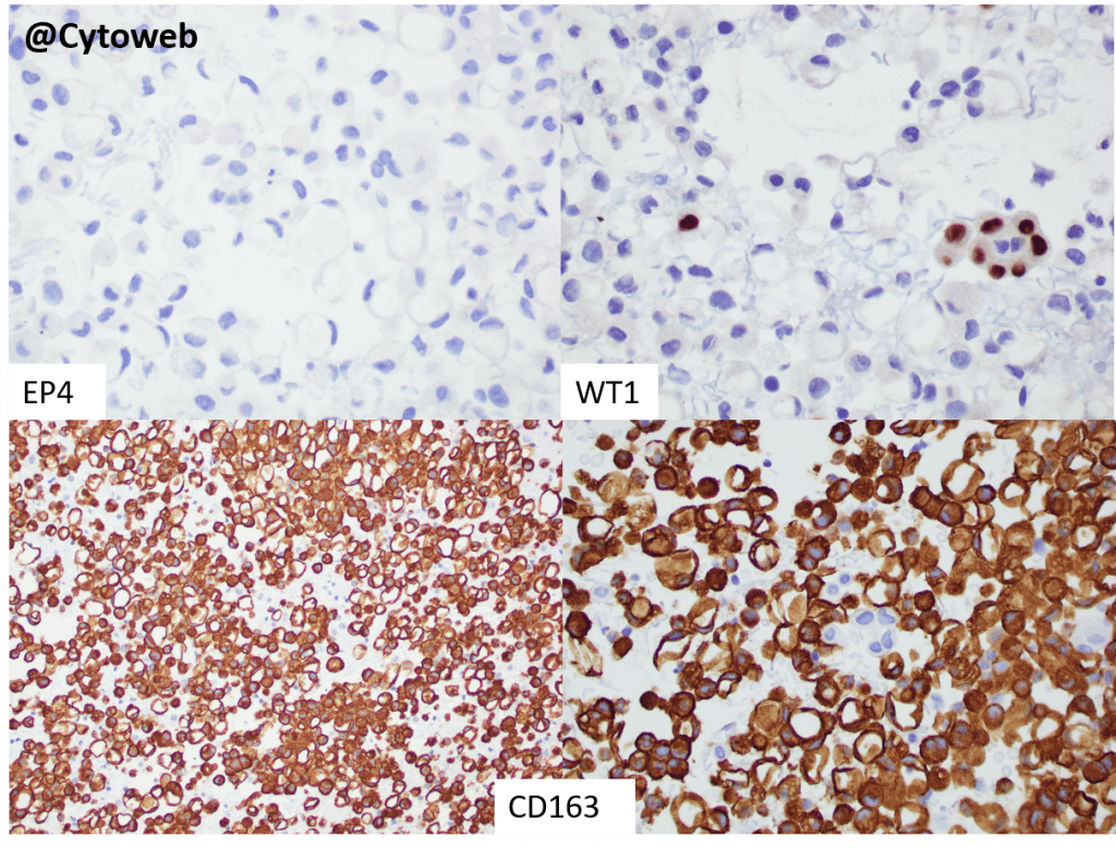
The cells with signet ring morphology are negative for the epithelial marker EP4 and the mesothelial marker WT1. A small group of WT1-positive mesothelial cells is seen. CD163 highlights numerous cells with signet ring cell morphology.
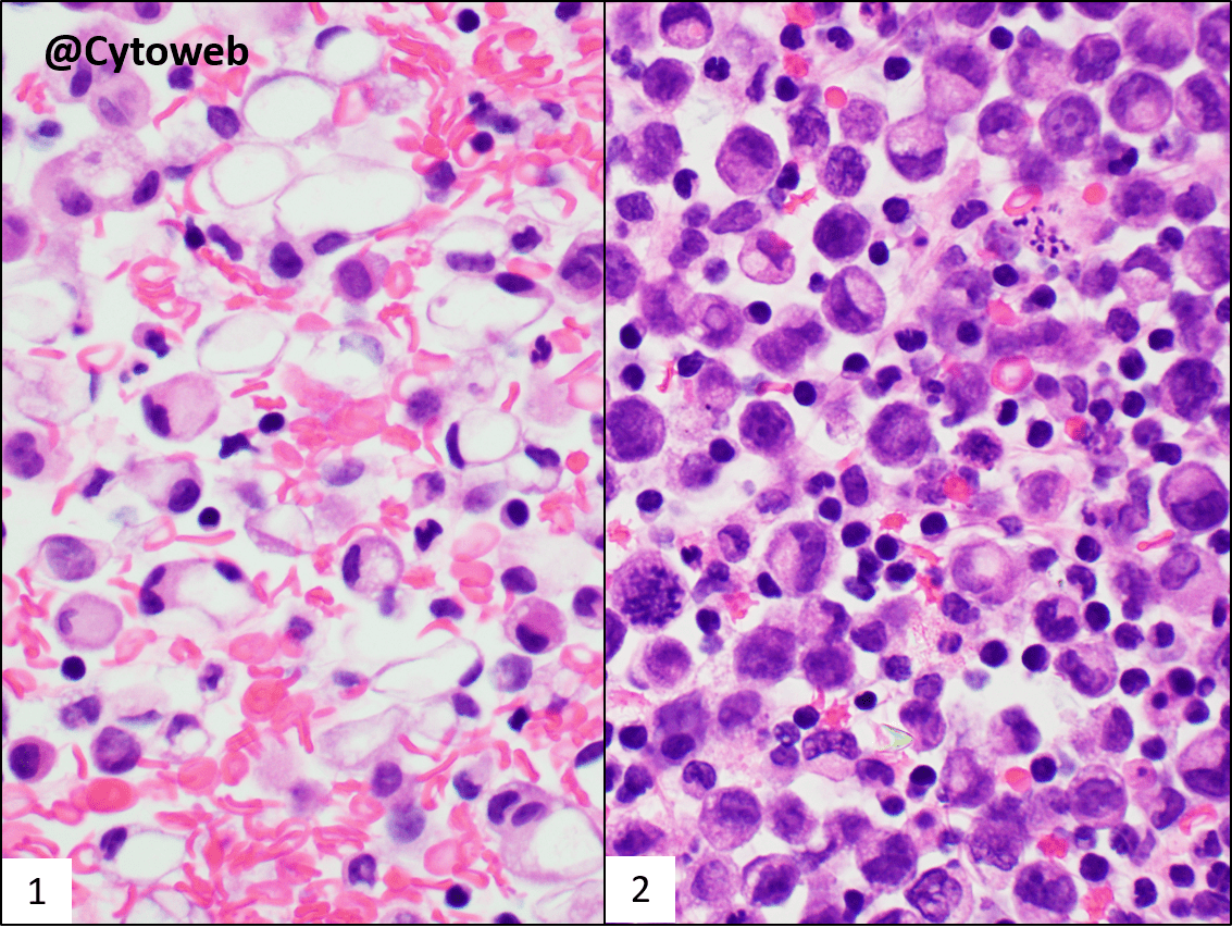
In body cavity fluid samples, the differential diagnoses for cells with signet ring morphology include:
- Metastatic adenocarcinoma with signet ring cell morphology
- Macrophages
- Mesothelial cells
The most helpful morphological features are the nuclei – significant nuclear abnormalities can be seen in adenocarcinoma (hyperchromatic or irregular nuclei with prominent nucleoli) as well as the nature of the cytoplasm (finely bubbly, pinkish in alcohol-stained smears, or containing a small mucin droplet in adenocarcinoma). When the cells contain water as a result of degenerative changes (e.g. in macrophages and mesothelial cells), the vacuole appears clear and empty as seen in this case.
A panel of immunostains will provide the answer as shown above.